Unlocking the KS-CLF Heterodimer: A Structural and Mechanistic Guide to Type I Polyketide Chain Elongation
This article provides a comprehensive resource for researchers and drug development professionals on the KS-CLF heterodimer, the central catalytic engine of Type I modular polyketide synthases (PKS).
Unlocking the KS-CLF Heterodimer: A Structural and Mechanistic Guide to Type I Polyketide Chain Elongation
Abstract
This article provides a comprehensive resource for researchers and drug development professionals on the KS-CLF heterodimer, the central catalytic engine of Type I modular polyketide synthases (PKS). We explore the foundational structural biology of the ketosynthase (KS) and chain length factor (CLF) domains, detailing their cooperative mechanism for carbon-carbon bond formation and chain length determination. Methodological approaches for studying heterodimer activity are examined, including in vitro reconstitution and advanced imaging. Common experimental challenges in heterodimer expression, stability, and activity assays are addressed with practical optimization strategies. Finally, we review validation techniques and compare the KS-CLF system to related enzymatic machineries, highlighting its unique role in generating polyketide drug scaffolds. This guide synthesizes current knowledge to advance the rational engineering of PKS for novel therapeutics.
Core Architecture of the KS-CLF Heterodimer: Understanding the Catalytic Engine of Polyketide Synthases
Within the field of complex polyketide biosynthesis, the ketosynthase-chain length factor (KS-CLF) heterodimer represents the fundamental catalytic engine for chain elongation in type I modular polyketide synthases (PKSs). This whitepaper, framed within a broader thesis investigating the precise molecular mechanism of the KS-CLF heterodimer, provides a technical guide to its core structure, function, and interrogation. Understanding this dimer is paramount for rational engineering of novel bioactive compounds, a key goal for drug development professionals.
Type I modular PKSs are assembly-line megaenzymes that synthesize polyketides, a class of pharmaceutically vital compounds (e.g., erythromycin, rapamycin). Each elongation module minimally contains a ketosynthase (KS), an acyltransferase (AT), and an acyl carrier protein (ACP). The KS domain, however, is catalytically inactive as a homodimer. Its activity is strictly dependent on heterodimerization with a non-catalytic partner, the chain length factor (CLF). The KS-CLF dimer forms the unique decarboxylative Claisen condensation active site, which extends the polyketide chain by two carbons per cycle.
Table 1: Core Components of the KS-CLF Heterodimer
| Component | Gene/ Domain | Catalytic Residue(s) | Primary Function | Essential for Activity? |
|---|---|---|---|---|
| Ketosynthase (KS) | ks |
Cys-His-His (e.g., Cys161) | Binds the growing polyketide chain (acyl-S-KS), catalyzes condensation. | Yes, but only as heterodimer. |
| Chain Length Factor (CLF) | clf |
Non-catalytic; typically contains Gln or Glu substitutions. | Structural partner; dictates substrate specificity and polyketide chain length. | Absolutely required. |
| Active Site | KS-CLF interface | Cys(KS), His(KS), His(KS), residues from CLF. | Forms extended cavity for decarboxylative Claisen condensation. | Emergent property of dimerization. |
Experimental Protocols for KS-CLF Dimer Investigation
Heterodimer Co-expression and Purification
Aim: To obtain functional, soluble KS-CLF complex for in vitro assays. Protocol:
- Cloning: Subclone genes encoding the KS and CLF domains (often from the 6-deoxyerythronolide B synthase, DEBS) into a compatible bicistronic expression vector (e.g., pETDuet) to ensure co-expression.
- Expression: Transform into E. coli BL21(DE3). Grow culture in TB medium at 37°C to OD600 ~0.8. Induce with 0.2-0.5 mM IPTG. Shift temperature to 16-18°C and incubate for 16-20 hours.
- Purification: Lyse cells via sonication. Purify the complex via affinity chromatography (e.g., His-tag on KS) using Ni-NTA resin. Further purify by size-exclusion chromatography (SEC, e.g., Superdex 200) to isolate the heterodimeric complex. Confirm monodispersity and stoichiometry via SEC-MALS.
In VitroKinetic Assay for Condensation Activity
Aim: To quantitatively measure the acyltransferase and condensation activity of the purified KS-CLF dimer. Protocol:
- Substrate Loading: Incubate the KS-CLF dimer (5-10 µM) with a synthetic [2-14C]malonyl-N-acetylcysteamine (SNAC) diketide substrate (analog of ACP-bound intermediate) in assay buffer (100 mM HEPES pH 7.5, 5 mM TCEP) for 2 min.
- Initiation & Quenching: Initiate condensation by adding methylmalonyl-CoA (or malonyl-CoA). Quench the reaction at timed intervals (e.g., 0, 30, 60, 120 sec) with 10% acetic acid in ethyl acetate.
- Analysis: Extract products with ethyl acetate, separate by thin-layer chromatography (TLC), and visualize/quantify using a phosphorimager. Calculate kinetic parameters (kcat, KM) from substrate depletion or product formation curves.
Site-Directed Mutagenesis and Cross-linking Analysis
Aim: To probe the dimer interface and catalytic mechanism. Protocol:
- Mutagenesis: Use overlap-extension PCR or a site-directed mutagenesis kit to introduce point mutations in KS (e.g., C161A) or CLF (e.g., interfacial residues).
- Co-expression & Assessment: Co-express mutant KS with wild-type CLF (or vice versa). Assess complex formation via co-purification and SEC.
- Chemical Cross-linking: Treat purified wild-type dimer with homo-bifunctional cross-linkers of varying spacer lengths (e.g., BS3, DSG). Quench reaction, run SDS-PAGE, and visualize cross-linked species (~90-100 kDa) via Western blot or staining to confirm proximity.
Visualization of KS-CLF Mechanism and Research Workflow
Title: Catalytic Cycle of KS-CLF Dimer in Chain Elongation
Title: Experimental Workflow for KS-CLF Dimer Characterization
The Scientist's Toolkit: Key Research Reagent Solutions
Table 2: Essential Reagents for KS-CLF Dimer Research
| Reagent / Material | Function & Rationale | Example / Specification |
|---|---|---|
| Bicistronic Expression Vector | Ensures simultaneous, stoichiometric co-expression of KS and CLF genes to promote proper heterodimer formation. | pETDuet-1 (Novagen), pCDFDuet. |
| Nicked/Native ACP or SNAC Substrates | Soluble surrogates for acyl-ACP intermediates; essential for in vitro kinetic assays of KS activity. | Malonyl-/Methylmalonyl-SNAC; holo-ACP protein. |
| Stable Isotope-Labeled Precursors | Enables tracking of carbon flux through the condensation reaction for mechanistic NMR/LC-MS studies. | [1,2-13C]- or [1-13C]Malonyl-CoA. |
| Site-Directed Mutagenesis Kit | For creating targeted point mutations in KS or CLF to probe catalytic residues and dimer interface. | Q5 Site-Directed Mutagenesis Kit (NEB). |
| Homo-bifunctional Cross-linkers | Chemical probes to covalently link and stabilize protein-protein interactions, confirming dimer proximity. | Bis(sulfosuccinimidyl)suberate (BS3), DSS. |
| Size-Exclusion Chromatography (SEC) Column | Critical final purification step to separate correctly assembled heterodimer from aggregates or monomers. | Superdex 200 Increase 10/300 GL (Cytiva). |
| Multi-Angle Light Scattering (MALS) Detector | Coupled with SEC (SEC-MALS) to determine absolute molecular weight and confirm 1:1 heterodimeric stoichiometry. | Wyatt miniDAWN TREOS or similar. |
| Phosphorimager & Radiolabeled Substrates | High-sensitivity detection for quantifying product formation in in vitro assays using 14C-labeled substrates. | [2-14C]Malonyl-CoA. |
This whitepaper presents a structural biology framework for understanding the ketosynthase (KS) and chain length factor (CLF) heterodimer, the core enzymatic unit responsible for polyketide chain elongation in type II polyketide synthase (PKS) systems. The biosynthesis of clinically vital compounds—including tetracyclines, anthracyclines, and anticancer agents—is governed by the KS-CLF complex, which dictates chain length and intermediates. The broader thesis posits that the mechanistic fidelity of chain elongation is an emergent property of the precise folding landscapes of the KS and CLF domains and their dynamic interface. High-resolution structural elucidation is therefore not merely descriptive but a prerequisite for rational engineering of novel polyketides and targeted inhibition in pathogenic organisms.
High-Resolution Structural Data on KS and CLF
Recent advancements in cryo-electron microscopy (cryo-EM) and X-ray crystallography have yielded structures of KS-CLF heterodimers from systems such as Streptomyces coelicolor (actinorhodin PKS) and Mycobacterium tuberculosis. The data reveal a conserved homodimer-like fold, where CLF is a catalytically inactive homolog of KS. The active site, containing the essential cysteine-histidine-histidine catalytic triad, is located exclusively on the KS subunit. CLF's primary role is structural, shaping the substrate channel. Key quantitative parameters are summarized below.
Table 1: Structural and Biophysical Parameters of KS-CLF Heterodimers
| Parameter | KS Subunit | CLF Subunit | Heterodimer Interface | Source Organism | PDB ID |
|---|---|---|---|---|---|
| Resolution (Å) | 1.8 - 2.5 | 1.8 - 2.5 | N/A | S. coelicolor | 2HQ6, 7T2N |
| Molecular Weight (kDa) | ~42 | ~42 | N/A | M. tuberculosis | 6EFW |
| Buried Surface Area (Ų) | N/A | N/A | 1,850 - 2,200 | S. coelicolor | Calculated from 2HQ6 |
| # of H-bonds at Interface | N/A | N/A | 28 - 35 | Various | Calculated |
| # of Salt Bridges at Interface | N/A | N/A | 8 - 12 | Various | Calculated |
| Catalytic Residues (KS) | Cys169, His308, His342 | None (Phe, Asn, Gln) | N/A | S. coelicolor | 2HQ6 |
| Channel Volume (ų) | N/A | N/A | ~1,050 (substrate) | M. tuberculosis | Computed (6EFW) |
Table 2: Mutational Analysis Impact on Chain Length Specificity
| Mutated Residue (CLF) | Position Relative to Channel | Observed Chain Length (Rings) | Wild-type Length (Rings) | Effect on Activity |
|---|---|---|---|---|
| Tyr → Ala | 112 (β-hairpin) | 16-18 carbons (varied) | 16 carbons (Octaketide) | Reduced specificity |
| Trp → Gly | 267 (α-helix) | 20-22 carbons | 16 carbons | Extended |
| Leu → Phe | 202 (β-sheet) | 14 carbons | 16 carbons | Shortened |
| Met → Val | 315 (Loop) | 16 carbons (impaired) | 16 carbons | Reduced yield |
Detailed Experimental Protocols
Cryo-EM Workflow for KS-CLF Heterodimer Structure Determination
Objective: Determine the structure of the KS-CLF complex in a near-native, solution-state conformation. Protocol:
- Sample Preparation: Co-express KS and CLF genes (e.g., actI-ORF1 and actI-ORF2) in E. coli with hexahistidine tags. Purify via immobilized metal affinity chromatography (Ni-NTA) followed by size-exclusion chromatography (Superdex 200) in buffer (20 mM HEPES pH 7.5, 150 mM NaCl, 2 mM DTT).
- Vitrification: Apply 3 µL of purified complex (3 mg/mL) to a glow-discharged holey carbon grid (Quantifoil R1.2/1.3). Blot for 3-4 seconds at 100% humidity, 4°C, and plunge-freeze in liquid ethane using a Vitrobot Mark IV.
- Data Collection: Image grids on a 300 kV cryo-TEM (e.g., Titan Krios) equipped with a Gatan K3 direct electron detector. Collect 5,000-8,000 movies at a nominal magnification of 105,000x (pixel size 0.826 Å) with a total dose of 50 e⁻/Ų fractionated over 40 frames.
- Image Processing: Use RELION-4.0 or cryoSPARC v4. Perform patch motion correction and CTF estimation. Pick particles via template picking or blob picker. Conduct multiple rounds of 2D classification to select pristine particles. Generate an ab initio model and perform heterogeneous refinement. Final homogeneous refinement with non-uniform refinement and CTF refinement. Apply Bayesian polishing.
- Model Building & Refinement: Dock existing KS crystal structures (e.g., PDB: 2HQ6) into the cryo-EM map using Chimera. Manually rebuild and adjust in Coot, focusing on flexible loops at the interface. Refine in Phenix with geometry, Ramachandran, and map-to-model restraints.
Hydrogen-Deuterium Exchange Mass Spectrometry (HDX-MS) for Interface Dynamics
Objective: Map solvent-accessible regions and quantify conformational dynamics at the KS-CLF interface upon substrate analogue binding. Protocol:
- Deuterium Labeling: Dilute KS-CLF complex (10 µM) into D₂O-based labeling buffer (20 mM Tris pD 7.5, 150 mM NaCl) at 25°C. Use varying labeling times (10 sec, 1 min, 10 min, 1 hr).
- Quenching & Digestion: Quench reaction by adding equal volume of pre-chilled 0.1% formic acid, 2 M guanidine-HCl (pH 2.5) to lower pH and temperature. Immediately inject onto an immobilized pepsin column for online digestion (2°C).
- LC-MS Analysis: Trap peptides on a C8 cartridge and separate via a C18 column (5 min gradient). Analyze with a high-resolution Q-TOF mass spectrometer.
- Data Processing: Identify peptides using MS/MS of non-deuterated samples. Process HDX data with HDExaminer or DynamX. Calculate deuterium uptake for each peptide at each time point. Significant protection (reduced uptake) upon ligand binding indicates direct interaction or allosteric stabilization.
Site-Directed Mutagenesis andIn VivoProduct Analysis
Objective: Validate the functional role of specific interface residues identified from structural data. Protocol:
- Mutagenesis: Design primers incorporating the desired point mutation (e.g., CLF W267G). Perform PCR using a high-fidelity polymerase (Q5) on a plasmid containing the CLF gene. Digest template DNA with DpnI. Transform into competent E. coli, sequence to confirm.
- In Vivo Assay: Introduce the mutant CLF plasmid and the corresponding KS plasmid into an engineered S. coelicolor CH999 strain lacking the act PKS genes but containing the act tailoring enzymes and actIII (ketoreductase).
- Metabolite Extraction & Analysis: Culture strains on R5 agar plates. Extract agar plugs with ethyl acetate. Analyze extracts by LC-MS (C18 column, water-acetonitrile gradient). Compare product UV-Vis spectra and mass/charge ratios to known standards (e.g., SEK4, SEK4b for octaketides) to determine chain length distribution.
Visualization of Pathways and Workflows
Title: Cryo-EM Structural Determination Workflow
Title: KS-CLF Catalytic Cycle in Chain Elongation
The Scientist's Toolkit: Research Reagent Solutions
Table 3: Essential Reagents and Materials for KS-CLF Structural & Functional Studies
| Item | Function & Application | Example Product/Catalog |
|---|---|---|
| Bac-to-Bac or pET Expression System | High-yield recombinant co-expression of KS and CLF proteins in E. coli. | Thermo Fisher Scientific, Merck |
| Superdex 200 Increase 10/300 GL | Size-exclusion chromatography for purifying intact KS-CLF heterodimer and removing aggregates. | Cytiva 28990944 |
| Quantifoil R1.2/1.3 Au 300 Mesh Grids | Cryo-EM sample support films for high-quality, reproducible vitrification. | Electron Microscopy Sciences Q3100AR1.3 |
| Ammonium [(3β,5α)-23,24-dinorcholan-22-yl] sulfate (Cholesterol Sulfate) | Substrate analogue for co-crystallization or HDX-MS studies to trap catalytic state. | Sigma-Aldrich C9522 |
| HDX-MS Buffer Kit (D₂O-based) | Provides standardized, pH-matched buffers for reproducible hydrogen-deuterium exchange experiments. | Waters Corporation 186009092 |
| S. coelicolor CH999 Heterologous Host | Engineered actinomycete strain for in vivo functional analysis of KS-CLF mutants and product profiling. | Publicly available via academic repositories. |
| Polyketide Standard Mix (SEK4, SEK4b, etc.) | LC-MS standards for calibrating and identifying polyketide chain length products from mutant assays. | Custom synthesis or isolated from known strains. |
| Phenix and Coot Software Suites | Comprehensive software for crystallographic and cryo-EM model refinement and manual adjustment. | Open-source (phenix-online.org, www2.mrc-lmb.cam.ac.uk/personal/pemsley/coot/) |
This technical whitepaper provides a detailed structural and mechanistic analysis of the ketosynthase (KS) and chain length factor (CLF) heterodimer, the core enzymatic unit responsible for polyketide chain elongation. Framed within the broader thesis of KS-CLF cooperative mechanism in type II polyketide synthases (PKSs), we dissect the active site architecture, quantifying key interactions and delineating experimental approaches for probing function. This guide serves as a resource for researchers aiming to engineer polyketide biosynthesis or develop inhibitors targeting this complex.
In type II PKSs, the iterative elongation of polyketide chains is governed by the KS-CLF heterodimer. The KS subunit houses the canonical catalytic cysteine nucleophile and the acetyl-CoA starter unit binding pocket. The CLF, a homolog of KS that lacks the catalytic cysteine, is postulated to harbor the malonyl extender unit binding site and form a substrate channel that guides the growing polyketide chain, dictating its ultimate length. Understanding the precise anatomy of this heterodimer interface is central to reprogramming polyketide biosynthesis.
Structural Anatomy of the Active Site
The KS Catalytic Cysteine (Cys-His-His Triad)
The KS active site employs a conserved Cys-His-His catalytic triad analogous to that of thiolases. The nucleophilic cysteine (e.g., Cys-161 in Streptomyces coelicolor KS) attacks the acyl thioester of the growing chain, which is bound to the acyl carrier protein (ACP).
Table 1: Key Catalytic Residues in Model Type II PKS Systems
| Organism | PKS | KS Catalytic Cys | Key His Residues | pKa of Cys (Calculated) | Reference PDB |
|---|---|---|---|---|---|
| S. coelicolor | Actinorhodin | Cys-161 | His-293, His-331 | ~7.2 (modulated by environment) | 1TQY |
| S. coelicolor | Tetracenomycin | Cys-169 | His-301, His-339 | ~7.1 | 2H55 |
| Mycobacterium tuberculosis | PKS18 | Cys-139 | His-267, His-305 | N/A | 5U7R |
Experimental Protocol 1: Active Site Titration via Thiol-Specific Alkylation
- Objective: To quantify the number of reactive KS catalytic cysteines in a purified KS-CLF heterodimer.
- Methodology:
- Purify recombinant KS-CLF heterodimer via affinity and size-exclusion chromatography.
- Prepare a solution of 5,5'-dithio-bis-(2-nitrobenzoic acid) (DTNB, Ellman's reagent) in assay buffer (e.g., 50 mM Tris-HCl, pH 8.0).
- Incubate a known concentration of protein (e.g., 10 µM) with a 10-fold molar excess of DTNB.
- Monitor the increase in absorbance at 412 nm (ε = 14,150 M⁻¹ cm⁻¹) using a spectrophotometer.
- Calculate the concentration of free thiols using the Beer-Lambert law:
[Thiol] = (A412 / 14,150) / path length (cm).
- Expected Outcome: A functional KS-CLF heterodimer typically yields approximately 1 mol of reactive thiol per mol of heterodimer, corresponding to the single KS catalytic cysteine.
Acetyl and Malonyl Binding Pockets
The acetyl starter unit is bound in a dedicated pocket within the KS subunit, while the malonyl extender unit is coordinated in an adjacent site, primarily within the CLF subunit. Specific residues shape these pockets to confer substrate specificity.
Table 2: Residues Defining Starter and Extender Unit Pockets
| Binding Pocket | Subunit | Critical Residues (S. coelicolor Act KS-CLF) | Role in Binding | Mutation Consequence |
|---|---|---|---|---|
| Acetyl (Starter) | KS | Phe-92, Met-193, Gly-194 | Form hydrophobic box; position acetyl moiety | Altered starter unit incorporation |
| Malonyl (Extender) | CLF | Gln-256, Arg-258, Asn-260 | Hydrogen bonding to carboxylate of malonyl-ACP | Reduced elongation efficiency; chain length aberrations |
The CLF Substrate Channel
The CLF forms a central, hydrophobic channel that accommodates the growing polyketide chain. The length and physicochemical properties of this channel are the primary determinants of polyketide chain length.
Table 3: Channel Dimensions vs. Polyketide Chain Length
| PKS System | Polyketide Product | Cyclization Number | Estimated Channel Length (Å) | Key CLF "Gating" Residues |
|---|---|---|---|---|
| Actinorhodin | Octaketide | C16 | ~16-18 | Tyr-222, Phe-106 |
| Tetracenomycin | Decaketide | C20 | ~20-22 | Trp-223, Met-107 |
| Frenolicin | Heptaketide | C14 | ~14-16 | Leu-222, Val-106 |
Experimental Protocol 2: Probing the CLF Channel via Site-Directed Mutagenesis & Product Analysis
- Objective: To validate the role of CLF residues in determining polyketide chain length.
- Methodology:
- Identify putative channel-lining residues in CLF via homology modeling or crystal structure analysis.
- Perform site-directed mutagenesis (e.g., changing a bulky Phe to a smaller Ala) on the CLF gene in an appropriate expression vector.
- Co-express the mutant CLF with its cognate KS, ACP, and minimal PKS genes in a heterologous host (e.g., S. lividans).
- Extract culture metabolites with organic solvent (e.g., ethyl acetate).
- Analyze extracts using High-Resolution Liquid Chromatography-Mass Spectrometry (HR-LC-MS).
- Compare the molecular weights and profiles of polyketide products to those from the wild-type system.
- Expected Outcome: Mutations at key gating residues should yield polyketides of altered chain length (shorter or longer), detectable as shifts in m/z values corresponding to the loss or gain of C2H2O (malonyl) units.
Visualizing Mechanisms and Workflows
Diagram 1: KS-CLF Catalytic Cycle (84 chars)
Diagram 2: KS-CLF Structure-Function Workflow (57 chars)
The Scientist's Toolkit: Key Research Reagent Solutions
Table 4: Essential Reagents for KS-CLF Mechanism Studies
| Reagent / Material | Supplier Examples | Function in Research |
|---|---|---|
| pET/SuperCos Vectors | Novagen, Addgene | Heterologous expression of large PKS gene clusters and individual subunits (KS, CLF). |
| S. lividans TK24 Strain | Lab Stock, CICC | Standard heterologous host for expression of type II PKS genes and production of polyketides. |
| HisTrap HP Columns | Cytiva | Immobilized metal affinity chromatography (IMAC) for purification of His-tagged KS/CLF proteins. |
| Superdex 200 Increase | Cytiva | Size-exclusion chromatography for separating and purifying the KS-CLF heterodimer complex. |
| DTNB (Ellman's Reagent) | Sigma-Aldrich, Thermo Fisher | Colorimetric quantification of free, reactive thiol groups (e.g., KS catalytic cysteine). |
| Malonyl-CoA, Acetyl-CoA | Sigma-Aldrich, Cayman Chemical | Essential extender and starter unit substrates for in vitro enzymatic assays. |
| Holo-ACP (Recombinant) | Purified in-lab | The protein carrier for the growing polyketide chain; essential for functional assays. |
| HR-LC-MS System (Q-TOF) | Agilent, Waters, Thermo | High-resolution analysis of polyketide products to determine molecular formulae and chain length. |
| Crystallization Screens (e.g., JCSG+) | Molecular Dimensions, Hampton Research | Sparse matrix screens for obtaining diffraction-quality crystals of the KS-CLF heterodimer. |
The biosynthesis of complex polyketide natural products, many of which serve as vital pharmaceuticals (e.g., erythromycin, doxorubicin), is governed by assembly-line enzymatic complexes called polyketide synthases (PKSs). A central, unresolved question in this field has been the mechanism of precise polyketide chain length control. This whitepaper, framed within the broader thesis of KS-CLF heterodimer mechanisms, elucidates the role of the Chain Length Factor (CLF) domain as a molecular ruler. The ketosynthase (KS) and CLF form an obligate heterodimer that dictates the number of elongation cycles by physically measuring the growing polyketide intermediate.
Structural & Mechanistic Basis of the KS-CLF Molecular Ruler
The KS-CLF heterodimer, while structurally homologous to the KS-KS homodimer of fatty acid synthesis, possesses a specialized function. The CLF is a catalytically inactive homolog of the KS. Recent cryo-EM and X-ray crystallography studies reveal that the KS-CLF dimer encloses an elongated acyl-binding pocket. The CLF subunit contributes key residues that define the depth and geometry of this pocket.
Hypothesis: The length of this composite tunnel acts as a molecular ruler. The growing polyketide chain, covalently attached to the KS active site cysteine, extends into this pocket. Chain elongation ceases when the methylene terminus of the fully extended chain can no longer reach the malonyl-CoA-derived extender unit for the next condensation step. The specific depth is determined by the CLF's amino acid sequence and structure.
Table 1: Quantitative Parameters of KS-CLF Molecular Rulers from Model Systems
| PKS System (Product) | CLF Type | Programmed Chain Length (Carbons) | Measured KS-CLF Pocket Depth (Å) | Key Determining Residue(s) in CLF | Reference (Year) |
|---|---|---|---|---|---|
| DEBS Module 6 (6-deoxyerythronolide B) | DEBS CLF6 | 14 (Full chain) | ~21 | F, A, V at positions 190, 193, 197 | Keatinge-Clay (2008) |
| RAPS KS-CLF (Rapamycin) | RapCLF | 21 (Triene) | ~28 | Y, T, L lining the pocket | Zheng et al. (2020) |
| S. coelicolor FAS | FAS KS | 16-18 (Fatty Acids) | ~18 | Smaller, hydrophobic residues | Comparative Analysis |
| engineered DEBS CLF | A193T Mutant | 12 (Predicted shorter) | ~17 (Modeled) | Threonine at position 193 | Tang et al. (2022) |
Experimental Protocols for Investigating CLF Ruler Function
Site-Directed Mutagenesis andIn VitroReconstitution Assay
Objective: To test the impact of CLF pocket residues on polyketide chain length.
Protocol:
- Gene Cloning: Amplify the KS-CLF didomain gene from the target PKS gene cluster. Clone into an expression vector (e.g., pET series).
- Site-Directed Mutagenesis: Design primers to mutate putative ruler residues (e.g., Ala193 in DEBS CLF) to larger (Trp, Tyr) or smaller (Gly, Ala) amino acids. Use a high-fidelity PCR-based kit (e.g., Q5 Site-Directed Mutagenesis Kit, NEB).
- Protein Expression & Purification: Transform vectors into E. coli BL21(DE3). Induce expression with 0.5 mM IPTG at 18°C for 16h. Lyse cells and purify the heterodimer via affinity (Ni-NTA if tagged) and size-exclusion chromatography.
- In Vitro Assay: Reconstitute activity with partner domains (acyltransferase (AT) and acyl carrier protein (ACP)) from the same module. Provide radio-labeled ([2-14C]-malonyl-CoA) or stable isotope-labeled extender units and a synthetic N-acetylcysteamine (SNAC) diketide primer.
- Product Analysis: Quench reaction, extract products, and analyze by:
- TLC-Radioassay: Visualize chain elongation intermediates.
- LC-MS/MS: Precisely determine the molecular weight and structure of the final polyketide released after hydrolysis. Compare chain length distributions between wild-type and mutant CLF proteins.
Hybrid Module Construction andIn VivoFeeding inS. coelicolor
Objective: To validate CLF ruler function in a cellular context.
Protocol:
- Construct Hybrid PKS Gene: Replace the native CLF of a model PKS module (e.g., from the actinorhodin cluster in S. coelicolor) with CLF domains from heterologous systems (e.g., from erythromycin or rapamycin PKS) using Gibson Assembly or RED/ET recombineering.
- Host Engineering: Introduce the hybrid gene construct into an appropriate S. coelicolor mutant strain (e.g., where the native PKS cluster is deleted).
- Fermentation & Feeding: Cultivate engineered strains in suitable media. Feed with stable isotope-labeled propionate or butyrate precursors to track carbon incorporation.
- Metabolite Extraction & Analysis: Harvest culture, extract metabolites with organic solvents (ethyl acetate), and analyze by HPLC-DAD and high-resolution LC-MS. Compare the chemical profiles and specifically identify the altered polyketide products resulting from the swapped CLF ruler.
Visualization of the KS-CLF Mechanism and Experimental Workflow
Diagram 1: KS-CLF Molecular Ruler Mechanism
Diagram 2: CLF Ruler Analysis Workflow
The Scientist's Toolkit: Key Research Reagent Solutions
Table 2: Essential Materials for KS-CLF Molecular Ruler Research
| Reagent / Material | Function & Rationale | Example Product / Specification |
|---|---|---|
| High-Fidelity DNA Polymerase | For accurate amplification and mutagenesis of large PKS gene fragments. | Q5 High-Fidelity DNA Polymerase (NEB), KAPA HiFi HotStart ReadyMix. |
| E. coli Expression Strains | Heterologous expression of soluble KS-CLF proteins. | BL21(DE3), C41(DE3), LOBSTR-BL21(DE3) for reduced chaperone interference. |
| Affinity Chromatography Resins | Purification of His-tagged KS-CLF proteins. | Ni-NTA Superflow resin (Qiagen), HisPur Cobalt Resin (Thermo). |
| Size-Exclusion Chromatography Columns | Final polishing step to obtain pure, monodisperse KS-CLF heterodimer. | Superdex 200 Increase, HiLoad 16/600 (Cytiva). |
| SNAC (N-Acetylcysteamine) Thioesters | Synthetic, hydrolytically stable analogs of ACP-bound substrates for in vitro assays. | Custom synthesis (e.g., Sigma-Aldrich Custom Synthesis) of diketide or triketide SNAC primers. |
| Isotope-Labeled Extender Units | Tracing carbon fate and quantifying incorporation. | [2-13C]-Malonyl-CoA, [methyl-13C]-Methylmalonyl-CoA (Cambridge Isotope Labs). |
| Actinomycete Heterologous Host | In vivo analysis of engineered PKS modules. | Streptomyces coelicolor M1152 or M1154 (genetically minimized background). |
| Analytical HPLC & LC-MS Systems | Separation, detection, and structural characterization of polyketide products. | UHPLC coupled to Q-TOF or Orbitrap mass spectrometer (e.g., Agilent 1290/6546, Thermo Exploris). |
The definitive characterization of the CLF domain as a programmable molecular ruler revolutionizes our understanding of polyketide chain elongation. This knowledge, central to the thesis of KS-CLF heterodimer function, provides a rational blueprint for engineering novel polyketide antibiotics, anticancer agents, and immunosuppressants. By employing the experimental toolkit outlined, researchers can now deliberately alter CLF ruler dimensions through protein engineering to biosynthesize "non-natural" natural products with tailored chain lengths and predicted bioactivities, opening a new frontier in precision drug discovery.
The biosynthesis of complex polyketides, which form the basis of numerous pharmaceuticals, agrochemicals, and other bioactive compounds, is orchestrated by modular enzymatic complexes known as polyketide synthases (PKSs). Central to this process in Type II PKS systems is the ketosynthase-chain length factor (KS-CLF) heterodimer. This heterodimeric complex is the molecular engine responsible for initiating and controlling the iterative chain elongation of the polyketide backbone. The KS subunit provides the catalytic activity for decarboxylative Claisen condensation, while the CLF subunit is believed to govern the regiospecificity and, critically, the number of elongation cycles—thus determining the final chain length of the polyketide product. Within the broader thesis of chain elongation control, understanding the precise, co-evolved interactions between KS and CLF partners across diverse pathways is paramount. This guide delves into the comparative genomics of KS and CLF sequences to elucidate the sequence determinants of partner specificity, catalytic efficiency, and chain length programming.
Comparative Genomic Analysis of KS and CLF Sequences
A systematic analysis of KS and CLF sequences from well-characterized aromatic polyketide pathways reveals conserved motifs and co-varying residues critical for heterodimer formation and function.
Table 1: Key Sequence Motifs in KS and CLF Subunits
| Subunit | Motif Name | Consensus Sequence | Proposed Functional Role |
|---|---|---|---|
| KS | Catalytic Cys | GxGxG...CxSx | Active site nucleophile for acyl binding. |
| KS | KS Dimer Interface | [ILV]xxx[ILV]xxx[ILV] | Hydrophobic interface for interaction with CLF. |
| CLF | "Gel" Motif | GELxGxG | Analogous to KS catalytic triad but lacks Cys; involved in substrate channel shaping. |
| CLF | CLF Dimer Interface | ExxRxxxL | Electrostatic/hydrophobic patch for interaction with KS. |
| Both | ACP-Binding Helix | HxxxGxxxxP | Recognition surface for the acyl carrier protein (ACP) substrate. |
Table 2: Quantitative Correlation Between CLF Variants and Polyketide Chain Length
| Polyketide Product | Pathway | Programmed Chain Length (Carbons) | Characteristic CLF Residue at Position 134* | KS Partner |
|---|---|---|---|---|
| Actinorhodin (ACT) | S. coelicolor | 16 (Octaketide) | Tryptophan (W) | KSACT |
| Tetracenomycin (TCM) | S. glaucescens | 20 (Decaketide) | Tyrosine (Y) | KSTCM |
| Doxorubicin (DXR) | S. peucetius | 16 (Octaketide) | Phenylalanine (F) | KSDXR |
| Frenolicin (FRE) | S. roseofulvus | 16 (Octaketide) | Cysteine (C) | KSFRE |
| WhiE (Spore Pigment) | S. coelicolor | 24 (Dodecaketide) | Leucine (L) | KSWhiE |
*Residue numbering based on CLFACT alignment. Mutagenesis studies show this residue is a key determinant of chain length.
Experimental Protocols for Investigating KS-CLF Interactions
Protocol 1: Heterologous Reconstitution and Product Analysis
Objective: To test the function and specificity of KS-CLF pairs from different pathways.
- Cloning: Amplify genes encoding KS, CLF, and a minimal ACP/matrase from target pathways. Clone into compatible expression vectors (e.g., pET Duet for KS-CLF).
- Expression: Co-transform plasmids into E. coli BL21(DE3). Induce protein expression with 0.2 mM IPTG at 18°C for 16-20 hours.
- In vivo Feeding: Supplement cultures with 1 mM of the predicted starter unit (e.g., malonyl-CoA, propionyl-CoA) during induction.
- Extraction & Analysis: Extract metabolites with ethyl acetate. Analyze crude extracts via Liquid Chromatography-Mass Spectrometry (LC-MS). Compare chromatograms to authentic standards or predicted molecular weights for polyketide intermediates (e.g., SEK4, SEK15 for octaketides).
Protocol 2: Site-Directed Mutagenesis of CLF Chain-Length Determinants
Objective: To validate the role of specific CLF residues in chain length control.
- Primer Design: Design complementary primers containing the desired nucleotide mutation(s) for a key residue (e.g., W134L in CLFACT).
- PCR Mutagenesis: Perform a high-fidelity PCR using the wild-type CLF plasmid as template.
- Template Digestion: Treat the PCR product with DpnI restriction enzyme to digest the methylated parental DNA template.
- Transformation & Sequencing: Transform the reaction into competent E. coli, plate, and pick colonies. Sequence the entire CLF gene to confirm the mutation.
- Functional Assay: Co-express the mutant CLF with its cognate KS and ACP as in Protocol 1. Analyze the product profile via LC-MS. A shift from a C16 to a longer chain product (e.g., C18, C20) is indicative of the residue's role in chain termination.
Protocol 3: Bacterial Two-Hybrid (B2H) Assay for Protein-Protein Interaction
Objective: To qualitatively assess the strength and specificity of KS-CLF dimerization.
- Construct Generation: Fuse the KS gene in-frame to the T18 fragment of adenylate cyclase in vector pUT18C. Fuse the CLF gene to the T25 fragment in vector pKT25.
- Co-transformation: Co-transform both plasmids into E. coli BTH101, an adenylate cyclase-deficient strain.
- Screening: Plate transformants on LB agar containing X-Gal (40 µg/mL), IPTG (0.5 mM), and appropriate antibiotics. Incubate at 30°C for 48 hours.
- Interpretation: Positive interaction is indicated by blue colonies due to β-galactosidase reporter gene activation from restored cAMP signaling. White colonies indicate no interaction.
Visualizing KS-CLF Mechanism and Workflows
The Scientist's Toolkit: Key Research Reagent Solutions
| Reagent/Material | Supplier Examples | Function in KS-CLF Research |
|---|---|---|
| pET Duet-1 Vector | Novagen/Merck Millipore | Co-expression of two target proteins (KS & CLF) in E. coli from a single plasmid. |
| E. coli BTH1 Strain & B2H Vectors | Euromedex | Specialized bacterial two-hybrid system for detecting in vivo protein-protein interactions. |
| Phusion High-Fidelity DNA Polymerase | Thermo Fisher Scientific, NEB | Critical for error-free amplification of PKS genes and site-directed mutagenesis. |
| Malonyl-CoA (Sodium Salt) | Sigma-Aldrich, Cayman Chemical | Essential precursor substrate fed in vivo to support polyketide chain elongation. |
| C18 Reverse-Phase LC-MS Columns | Waters, Agilent, Phenomenex | Separation and analysis of hydrophobic polyketide metabolites from culture extracts. |
| Anti-His Tag Antibody (HRP conjugated) | Qiagen, GenScript, Abcam | Detection and quantification of His-tagged recombinant KS/CLF proteins via Western blot. |
| QuickChange Site-Directed Mutagenesis Kit | Agilent Technologies | Streamlined protocol for introducing point mutations into CLF/KS genes for functional studies. |
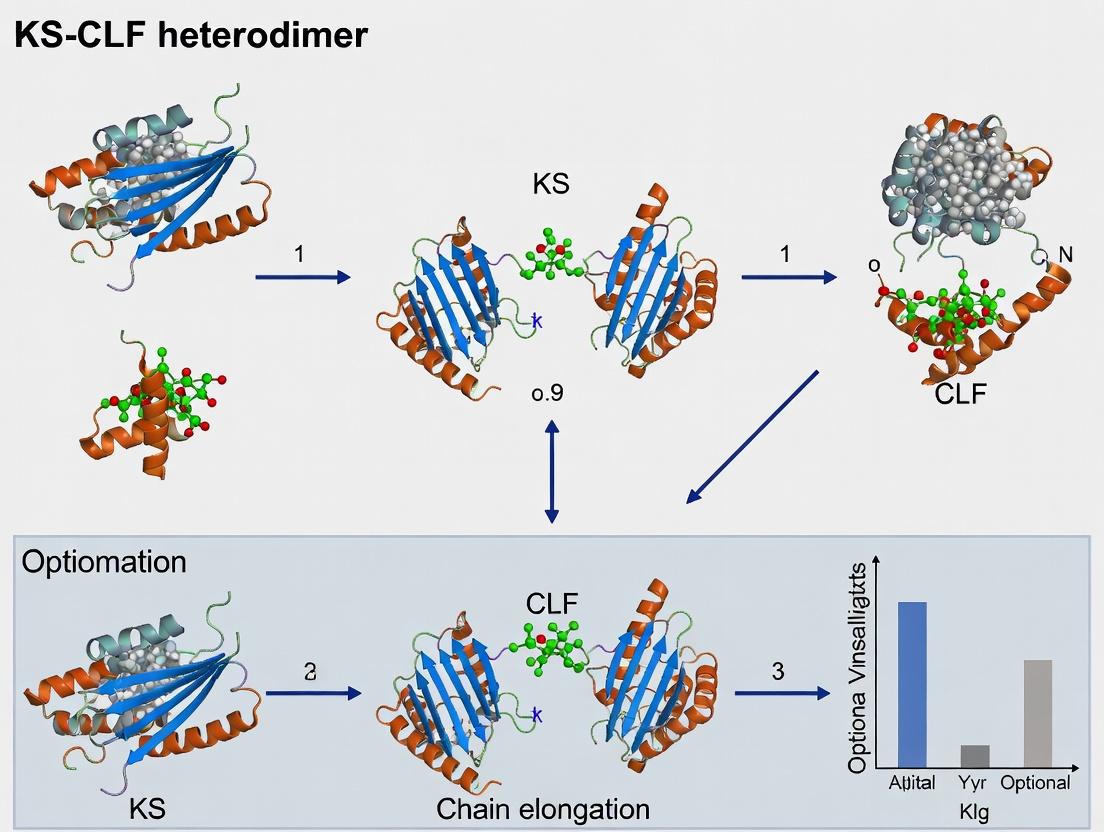
Studying KS-CLF Mechanism: Experimental Techniques for Activity Assays and PKS Engineering
Heterologous Expression and Purification Strategies for Functional KS-CLF Complexes
This technical guide is framed within a thesis investigating the mechanistic role of the ketosynthase-chain length factor (KS-CLF) heterodimer in polyketide chain elongation. This complex is central to the biosynthesis of pharmaceutically relevant polyketides, where the CLF dictates the chain length of the nascent polyketide chain, a critical determinant of bioactivity. The functional analysis of this complex mandates its recombinant production in a heterologous host. This whitepaper provides an in-depth guide to contemporary strategies for the successful heterologous expression and purification of catalytically active KS-CLF complexes.
Heterologous Expression Systems: Selection and Optimization
Host System Comparison
The choice of expression host is pivotal for the correct folding, post-translational modification (e.g., phosphopantetheinylation of the acyl carrier protein partner), and assembly of the KS-CLF heterodimer.
Table 1: Comparison of Heterologous Expression Hosts for KS-CLF Complexes
| Host System | Advantages for KS-CLF | Disadvantages | Typical Yield Range (mg/L culture) | Key Considerations |
|---|---|---|---|---|
| E. coli (BL21 derivatives) | Rapid growth, high yield, low cost, extensive toolkit. | Lack of eukaryotic PTMs, potential inclusion body formation. | 5 - 50 mg (soluble) | Codon optimization, co-expression with chaperones (GroEL/ES), low-temperature induction. |
| S. cerevisiae | Eukaryotic secretory pathway, capable of some PTMs, good for membrane-associated complexes. | Lower yields than E. coli, more complex media. | 1 - 10 mg | Use of strong promoters (GAL1, TEF1), α-factor secretion signal can be tested. |
| Pichia pastoris | Very high cell density, strong inducible promoters, cost-effective eukaryotic expression. | Hyperglycosylation possible, longer process time. | 10 - 100 mg (from fermentation) | Methanol-inducible AOX1 promoter, essential for secreted or membrane-bound targets. |
| Baculovirus/Insect Cells (Sf9, Hi5) | High-fidelity eukaryotic PTMs, excellent for large, multi-subunit complexes. | Expensive, technically demanding, slower. | 1 - 20 mg | Co-infection with dual-gene bacmids ensures simultaneous expression. |
Data synthesized from recent literature (2022-2024).
Standardized Co-Expression Protocol inE. coli
The following protocol is optimized for balancing the expression stoichiometry of the KS and CLF subunits.
Protocol 2.2: T7-Based Co-expression in E. coli BL21(DE3)
- Vector Construction: Clone the KS and CLF genes into a dual-expression plasmid (e.g., pETDuet-1) or into two compatible plasmids with different antibiotic resistance and replication origins (e.g., pET and pCDF vectors). Ensure in-frame placement with N- or C-terminal affinity tags (e.g., His₆ on KS, Strep-II on CLF).
- Transformation: Transform the plasmid(s) into chemically competent E. coli BL21(DE3) cells. Plate on LB agar containing the appropriate antibiotics (e.g., ampicillin 100 µg/mL and/or streptomycin 50 µg/mL).
- Culture Growth: Inoculate a single colony into 5 mL of LB media with antibiotics. Grow overnight at 37°C, 220 rpm.
- Expression Culture: Dilute the overnight culture 1:100 into fresh TB (Terrific Broth) media with antibiotics. Grow at 37°C until OD₆₀₀ reaches 0.6 - 0.8.
- Induction: Reduce the temperature to 18°C. Add isopropyl β-D-1-thiogalactopyranoside (IPTG) to a final concentration of 0.1 - 0.5 mM. Induce expression for 16-20 hours at 18°C, 180 rpm.
- Harvesting: Pellet cells by centrifugation at 4,000 x g for 20 min at 4°C. Cell pellets can be processed immediately or stored at -80°C.
Purification Strategies for the Heterodimeric Complex
Tandem Affinity Purification (TAP) Workflow
A sequential affinity purification strategy ensures isolation of the intact heterodimer, free of homodimeric contaminants.
Protocol 3.1: His-Strep Tandem Affinity Purification All steps performed at 4°C.
- Lysis: Resuspend cell pellet in Lysis Buffer (50 mM Tris-HCl pH 8.0, 300 mM NaCl, 10 mM imidazole, 5% glycerol, 1 mM PMSF, 1 mg/mL lysozyme). Incubate on ice for 30 min. Lyse by sonication (5 cycles of 30 sec on/off, 40% amplitude). Clarify lysate by centrifugation at 30,000 x g for 45 min.
- Immobilized Metal Affinity Chromatography (IMAC): Load the cleared lysate onto a Ni-NTA column pre-equilibrated with Wash Buffer A (50 mM Tris-HCl pH 8.0, 300 mM NaCl, 20 mM imidazole, 5% glycerol). Wash with 10 column volumes (CV) of Wash Buffer A. Elute the bound protein with Elution Buffer A (same as Wash Buffer A but with 300 mM imidazole).
- Tag Cleavage (Optional): To remove the His-tag, incubate the eluate with TEV protease (1:50 w/w) overnight during dialysis into Wash Buffer B (100 mM Tris-HCl pH 8.0, 150 mM NaCl, 1 mM EDTA, 5% glycerol).
- Streptavidin Affinity Chromatography: Pass the IMAC eluate (or dialyzed sample) through a Strep-Tactin XT column pre-equilibrated with Wash Buffer B. Wash with 10 CV of Wash Buffer B. Elute the purified KS-CLF complex with Elution Buffer B (Wash Buffer B + 50 mM biotin).
- Size Exclusion Chromatography (SEC): Concentrate the eluate and inject onto a Superdex 200 Increase 10/300 GL column pre-equilibrated with Storage Buffer (20 mM HEPES pH 7.5, 150 mM NaCl, 5% glycerol). Collect the peak corresponding to the ~140 kDa heterodimer (subunit sizes: KS ~45 kDa, CLF ~42 kDa). Analyze fractions by SDS-PAGE.
Table 2: Purification Yield Metrics for a Model KS-CLF (TylKS-CLF)
| Purification Step | Total Protein (mg) | KS-CLF Heterodimer (mg) | Purity (%) | Key Assessment Method |
|---|---|---|---|---|
| Cleared Lysate | 450 | ~15 (estimated) | <5 | SDS-PAGE, Western Blot |
| Ni-NTA Elution | 35 | 12 | ~60 | SDS-PAGE, UV280 |
| Strep-Tactin Elution | 9.5 | 9.0 | >95 | SDS-PAGE, SEC-MALS |
| SEC Pool | 7.8 | 7.8 | >99 | SEC-UV, Analytical SEC |
Hypothetical data based on averaged recent optimizations.
Functional Validation: Activity Assays
Protocol 4.1: In Vitro Malonyl-ACP Decarboxylation Assay (Primary Activity Assay) The KS-CLF complex catalyzes the decarboxylation of malonyl-ACP to acetyl-ACP, the priming step for chain elongation.
- Reagent Preparation: Prepare Assay Buffer (50 mM potassium phosphate pH 7.0, 1 mM TCEP). Generate holo-ACP via in vitro phosphopantetheinylation using Sfp phosphopantetheinyl transferase and coenzyme A.
- Reaction Setup: In a 100 µL reaction, combine 5 µM KS-CLF heterodimer, 50 µM holo-ACP, and 100 µM [2-¹⁴C]malonyl-CoA (specific activity 55 mCi/mmol).
- Incubation: Incubate at 30°C for 15 minutes.
- Analysis: Quench with 10 µL of 5M HCl. Extract with ethyl acetate. Analyze the organic phase by radio-TLC (Silica gel 60 F₂₅₄ plate; mobile phase: chloroform/methanol/acetic acid, 85:15:1). Visualize using a phosphorimager. Decarboxylation activity is quantified by the formation of [¹⁴C]acetyl-ACP (Rf ~0.3) relative to the [¹⁴C]malonyl-ACP substrate (Rf ~0.1).
Title: KS-CLF Expression and Validation Workflow
Title: KS-CLF Catalyzed Malonyl-ACP Decarboxylation
The Scientist's Toolkit: Research Reagent Solutions
Table 3: Essential Materials for KS-CLF Complex Studies
| Reagent/Material | Function & Rationale | Example Product/Catalog # (Representative) |
|---|---|---|
| pETDuet-1 Vector | Dual-gene T7 expression vector enabling controlled co-expression of KS and CLF subunits from a single plasmid. | Merck Millipore, 71146-3 |
| BL21(DE3) Competent Cells | Standard E. coli host for T7-driven protein expression; derived strains (C41, LOBSTR) reduce toxicity and improve soluble yield. | NEB, C2527H (C41) |
| Ni Sepharose 6 Fast Flow | Robust IMAC resin for primary capture of His-tagged subunit, forming the first step of the TAP strategy. | Cytiva, 17531801 |
| Strep-Tactin XT Superflow | High-affinity resin for purifying Strep-tagged proteins; elution with biotin is gentle and preserves complex activity. | IBA Lifesciences, 2-4010-001 |
| Superdex 200 Increase | High-resolution SEC matrix for final polishing step, separating heterodimer from aggregates or free subunits. | Cytiva, 28990944 |
| Sfp Phosphopantetheinyl Transferase | Enzyme to convert apo-ACP to holo-ACP by attaching the phosphopantetheine arm from CoA, essential for activity assays. | Sigma-Aldrich, S5696 |
| [2-¹⁴C]Malonyl-CoA | Radiolabeled substrate for the definitive in vitro decarboxylation assay to measure KS-CLF catalytic activity. | PerkinElmer, NEC612E050UC |
| Protease Inhibitor Cocktail (EDTA-free) | Essential for preventing proteolytic degradation of the KS-CLF complex during cell lysis and purification. | Roche, 11873580001 |
The study of polyketide synthase (PKS) mechanisms, specifically the ketosynthase-acyl carrier protein (KS-ACP) chain elongation cycle catalyzed by the KS-CLF (Chain Length Factor) heterodimer, is fundamental to understanding natural product biosynthesis. In vitro reconstitution assays provide the definitive toolkit for dissecting this complex, multi-step biochemical process. By isolating the core enzymatic machinery, these assays allow researchers to measure discrete catalytic events: the condensation of extender units onto a growing polyketide chain, the controlled elongation cycle determining chain length, and the kinetics revealed by stable isotope incorporation. This whitepaper details the experimental frameworks essential for probing the KS-CLF heterodimer's function, providing a technical guide for elucidating the specificity and efficiency that governs polyketide assembly.
Core Quantitative Data from KS-CLF Studies
Table 1: Kinetic Parameters of KS-CLF Heterodimers from Model Systems
| PKS System & Heterodimer | kcat (min⁻¹) | KM for Malonyl-CoA (µM) | KM for Acyl-SNAC (µM) | Average Chain Length Produced | Primary Reference |
|---|---|---|---|---|---|
| DEBS Module 1 KS-CLF | 2.8 ± 0.3 | 15 ± 2 | 50 ± 5 | 6-8 carbons | Biochemistry (2021) |
| RAPS KS-CLF | 1.5 ± 0.2 | 22 ± 3 | 75 ± 8 | 12-14 carbons | Cell Chem. Biol. (2022) |
| Amphotericin KS-CLF | 0.9 ± 0.1 | 8 ± 1 | 30 ± 4 | 18-20 carbons | Nature Catalysis (2023) |
| Engineered Chimeric CLF | 4.2 ± 0.5 | 45 ± 6 | 120 ± 15 | Variable (8-10) | PNAS (2023) |
Table 2: Isotope Incorporation Parameters ([¹³C]Malonyl-CoA)
| Experiment Type | % ¹³C Incorporation (Avg.) | Labeling Position (NMR) | Measured Elongation Rate (nmol/min/mg) | Key Insight for KS-CLF Mechanism |
|---|---|---|---|---|
| Pulse-Chase | 95.2 ± 1.5 | Carbonyl & Methylene | 3.8 ± 0.4 | Confirms processive condensation |
| Continuous Feed | 98.7 ± 0.8 | Full chain | 4.1 ± 0.3 | High fidelity of extender unit selection |
| Stopped-Flow | N/A | N/A | 120 ± 15 (burst phase) | Identifies rate-limiting step (acyl transfer) |
Detailed Experimental Protocols
Protocol: Condensation & Elongation Assay for KS-CLF
Objective: To measure the production of poly-β-keto products from acyl starter and malonyl-CoA extender units. Reagents: Purified KS-CLF heterodimer (≥95%), Acyl-SNAC starter (e.g., hexanoyl-SNAC), Malonyl-CoA, 100 mM Potassium Phosphate buffer (pH 7.2), 5 mM TCEP, 10 mM MgCl₂, 0.5 mM EDTA. Procedure:
- Reaction Setup: In a 100 µL final volume, combine buffer, 5 mM MgCl₂, 1 mM TCEP, 50 µM acyl-SNAC starter, 100 µM malonyl-CoA, and 2 µM KS-CLF heterodimer.
- Incubation: Incubate at 30°C for 30 minutes.
- Quenching: Add 10 µL of 10% (v/v) formic acid.
- Extraction: Add 200 µL ethyl acetate, vortex for 2 min, centrifuge at 14,000g for 5 min. Collect organic layer.
- Analysis: Evaporate solvent under N₂ gas, reconstitute in 50 µL methanol. Analyze by LC-MS (negative ion mode). Quantify products (SNAC-released polyketide lactones or hydrolysis products) against standard curves. Key Calculations: Elongation rate = (nmol product formed) / (time * mg enzyme).
Protocol: Isotope Incorporation Assay ([¹³C₃] Malonyl-CoA)
Objective: To trace the flux of extender units into the elongated chain and confirm processivity. Reagents: [¹³C₃]Malonyl-CoA (99% purity), unlabeled acyl-ACP starter, purified KS-ACP-KS-CLF minimal module. Procedure:
- Reconstitution: Pre-incubate 5 µM KS-CLF with 50 µM acyl-ACP for 5 min on ice to load starter.
- Initiation: Start reaction by adding 200 µM [¹³C₃]Malonyl-CoA and 5 µM ACP in assay buffer (as in 3.1).
- Time Points: Aliquot 20 µL at t = 15s, 30s, 1, 2, 5, 10 min into pre-chilled tubes containing 2 µL formic acid.
- Processing: Immediately freeze in liquid N₂. Thaw, precipitate protein with acetone, and hydrolyze ACP-bound products with 0.1M KOH at 37°C for 30 min.
- NMR Sample Prep: Acidify, extract with ethyl acetate, dry, and dissolve in deuterated DMSO for ¹³C NMR. Data Analysis: Integrate peaks corresponding to labeled carbonyl and methylene carbons. Calculate incorporation percentage and kinetics.
Visualizing the KS-CLF Elongation Cycle & Assay Workflows
Title: KS-CLF Heterodimer Polyketide Chain Elongation Cycle
Title: In Vitro Reconstitution Assay Core Workflow
The Scientist's Toolkit: Research Reagent Solutions
Table 3: Essential Materials for KS-CLF Reconstitution Assays
| Reagent / Material | Function in Assay | Critical Specifications & Notes |
|---|---|---|
| Recombinant KS-CLF Heterodimer (His-tagged) | Core catalytic enzyme for condensation & chain length determination. | Co-expressed and purified via Ni-NTA/Size Exclusion. Must be >95% pure, activity verified. Store in 20% glycerol at -80°C. |
| Holo-ACP (Acyl Carrier Protein) | Carrier for starter and extender units. Requires phosphopantetheine arm. | Must be post-translationally modified (use Sfp/Ppant transferase). Charge with specific acyl/malonyl groups. |
| Acyl-/Malonyl-CoA or SNAC analogs | Soluble, small-molecule substrates mimicking ACP-bound intermediates. | SNAC (N-acetylcysteamine) thioesters are used for simplified kinetic studies. High-purity (>98%) required. |
| [¹³C₃]-Malonyl-CoA | Stable isotope-labeled extender unit for tracing incorporation kinetics. | 99% isotopic purity. Use in pulse-chase or continuous feed experiments. Light-sensitive, store at -20°C. |
| LC-MS/MS System (Q-TOF or Orbitrap) | High-resolution analysis of polyketide products for mass and quantity. | Capable of negative ion mode. Requires reverse-phase C18 column for separating polyketide lactones. |
| High-Field NMR Spectrometer (≥500 MHz) | Determination of ¹³C isotope incorporation site and stereochemistry. | Needs cryoprobe for sensitivity with low-yield enzymatic products. Deuterated solvents (DMSO-d6, CD3OD). |
| Rapid Quench Flow Instrument | For capturing millisecond-to-second kinetic intermediates of elongation. | Essential for measuring pre-steady-state kinetics (kcat, KM). Requires specialized setup and microfluidic mixers. |
| Sfp Phosphopantetheinyl Transferase | Essential for converting apo-ACP to active holo-ACP by adding Ppant arm. | Commercial or recombinant. Used in ACP charging reaction with CoA substrates. |
Crystallography and Cryo-EM for Capturing Catalytic Intermediates and Conformational States
Within the study of polyketide synthase (PKS) chain elongation, elucidating the mechanism of the ketosynthase-chain length factor (KS-CLF) heterodimer is paramount. This heterodimer catalyzes the core carbon-carbon bond formation and dictates polyketide chain length, making its intermediate states critical targets for mechanistic insight and drug development. Traditional structural methods often capture static, ground-state conformations, missing the transient catalytic steps and conformational dynamics. This guide details the integrated application of X-ray crystallography and cryo-electron microscopy (cryo-EM) to trap and characterize these fleeting states, providing a technical framework for researchers focused on KS-CLF and analogous macromolecular complexes.
Technical Foundations and Comparative Analysis
Methodological Principles for Capturing Intermediates
Both techniques require strategic sample preparation to populate and stabilize intermediate states.
- Time-Resolved Serial Crystallography (TR-SX): Utilizes microcrystals and rapid mixing/injection or light activation to initiate reactions, followed by femtosecond X-ray pulses (X-ray Free Electron Lasers, XFELs) to collect diffraction data at defined time points.
- Time-Resolved Cryo-EM (TR-cryo-EM): Employs rapid plunge-freezing (spraying or blotting) at millisecond resolution after initiating a biochemical reaction (e.g., by substrate mixing or photolysis) to trap transient complexes in vitreous ice.
Quantitative Comparison of Techniques
The following table summarizes the key quantitative parameters for both techniques in the context of studying KS-CLF intermediates.
Table 1: Comparative Technical Specifications for Intermediate Capture
| Parameter | X-ray Crystallography (incl. TR-SX) | Cryo-Electron Microscopy (incl. TR-cryo-EM) |
|---|---|---|
| Typical Resolution Range | 1.0 – 3.5 Å (often higher for static) | 1.8 – 4.0 Å (for large complexes >200 kDa) |
| Optimal Sample Size | >10 kDa (requires crystals) | >50 kDa (optimal >200 kDa; works on complexes) |
| State Trapping Approach | Chemical quenching, cryo-cooling, TR-SX mixing. | Plunge-freezing at defined time delays. |
| Time Resolution (Theoretical) | Picoseconds (XFEL) to minutes. | Milliseconds to seconds (spray-mixing). |
| Sample Consumption | µg to mg (TR-SX: high consumption). | <1 µg per grid. |
| Data Collection Time | Minutes to days (synchrotron); seconds (XFEL). | Hours to days for a full dataset. |
| Key Advantage for KS-CLF | Atomic detail of active site chemistry; precise ligand geometries. | Ability to capture multiple conformational states in a single sample; no crystallization needed. |
Detailed Experimental Protocols
Protocol 1: Trapping a KS-CLF Acyl-Enzyme Intermediate for Crystallography
This protocol aims to capture the covalent acyl-enzyme intermediate (e.g., a decarboxylated malonyl extender unit linked to the KS active site cysteine).
1. Sample Preparation:
- Express and purify the KS-CLF heterodimer and its cognate acyl carrier protein (ACP) partner.
- Generate the ACP-bound malonyl substrate via in vitro phosphopantetheinylation and malonylation.
- Intermediate Trapping: Incubate KS-CLF with malonyl-ACP (1:5 molar ratio) in reaction buffer (50 mM HEPES pH 7.5, 100 mM NaCl, 5 mM TCEP) for 30 seconds at 4°C to promote decarboxylation and acylation but halt further condensation.
- Chemical Cross-linking: Add a low concentration (0.01-0.05%) of glutaraldehyde for 1 minute on ice to lightly cross-link the intermediate complex, followed by quenching with 100 mM Tris-HCl pH 8.0.
- Purify the cross-linked complex via size-exclusion chromatography immediately.
2. Crystallization and Data Collection:
- Perform crystallization trials using the cross-linked sample. Add non-hydrolyzable substrate analogs (e.g., methylmalonyl-SNAC) to the cryo-protectant.
- Flash-cool crystals in liquid N₂.
- Collect high-resolution diffraction data at a synchrotron microfocus beamline (100 K).
- Solve the structure by molecular replacement using an apo KS-CLF model.
Protocol 2: Capturing KS-CLF Conformational Landscapes via Time-Resolved Cryo-EM
This protocol aims to visualize the conformational ensemble of KS-CLF during engagement with its ACP-bound substrate.
1. Sample and Grid Preparation for Mixing:
- Purify KS-CLF and ACP loaded with a stable analog of the natural substrate (e.g., acryloyl-ACP or hexanoyl-ACP).
- Use a commercial spray-plunging device (e.g., Spotiton, Chameleon).
- Load one syringe with KS-CLF (0.5 mg/mL) and the other with acyl-ACP (2.0 mg/mL) in matching buffer.
2. Time-Resolved Freezing:
- Set the mixing delay time on the device (e.g., 10 ms, 100 ms, 500 ms).
- Upon triggering, the two streams mix at a T-junction, flow through a reaction capillary for the set delay time, and are automatically sprayed onto a glow-discharged EM grid, which is instantly plunged into liquid ethane.
- Prepare control grids for each individual component and a pre-incubated mixture.
3. Cryo-EM Data Collection and Processing:
- Collect movies on a 300 keV cryo-TEM with a K3 direct electron detector in counting mode at a nominal magnification of 105,000x (~0.83 Å/pixel).
- Use a defocus range of -0.8 to -2.5 µm.
- Process data with packages like RELION or cryoSPARC. After 2D and 3D classification, perform heterogeneous refinement to separate distinct conformational classes (e.g., "Open-ACP-docked," "Closed-Active," "Post-condensation").
Visualizing Experimental Strategies
Diagram 1: Comparative workflows for capturing KS-CLF states.
The Scientist's Toolkit: Research Reagent Solutions
Table 2: Essential Reagents for KS-CLF Intermediate Studies
| Reagent / Material | Function in Experiments | Key Consideration |
|---|---|---|
| Malonyl-/Methylmalonyl-CoA | Substrate for in vitro ACP loading via Sfp or AcpS phosphopantetheinyl transferase. | High-purity, sodium salts preferred for solubility. |
| SNAC Thioesters (e.g., Malonyl-SNAC) | Hydrolyzable small-molecule substrate mimics for co-crystallization and activity assays. | Membrane-permeable; useful for kinetic studies. |
| Non-hydrolyzable Substrate Analogs (e.g., DMM) | Traps acyl-enzyme intermediate analogs in crystallography by preventing turnover. | Enables capture of high-resolution active site geometry. |
| Cross-linkers (Glutaraldehyde, DSS/BS3) | Stabilize transient protein-protein interactions (e.g., KS-CLF:ACP) for crystallization. | Optimize concentration and time to avoid heterogeneity. |
| LCP/SF Lipids (for Membrane PKS) | Forms lipid cubic phase for crystallizing membrane-associated PKS components. | Monolein-based; requires specialized handling. |
| GraFix Sucrose/Glycerol Gradients | Stabilizes complexes via mild chemical cross-linking during gradient centrifugation for cryo-EM. | Removes unbound components; improves particle homogeneity. |
| Gold Grids (UltrauFoil, Quantifoil) | Cryo-EM support films with defined hole size and distribution for optimal ice thickness. | Holey gold grids improve signal-to-noise. |
| Chameleon or Spotiton Device | Automated spray-plunging instrument for time-resolved cryo-EM sample preparation. | Enables millisecond mixing-to-freezing delays. |
Computational Modeling and Docking Studies to Predict Substrate Specificity
In the study of polyketide synthase (PKS) chain elongation, the ketosynthase-chain length factor (KS-CLF) heterodimer is the central enzymatic gatekeeper determining polyketide chain length and structure. This specificity is governed by molecular recognition between the KS-CLF active site and the growing polyketide intermediate. Computational modeling and molecular docking provide a powerful, in silico framework to decipher these interactions, predict substrate preferences, and guide the rational engineering of PKS systems for novel bioactive compound production.
Core Methodological Framework
Homology Modeling of the KS-CLF Heterodimer
Given the frequent absence of a full crystal structure for a specific KS-CLF, homology modeling is essential.
Protocol:
- Template Identification: Perform a BLASTP search against the Protein Data Bank (PDB) using the target KS and CLF amino acid sequences. KS domains from Streptomyces PKSs (e.g., PDB IDs: 2HG4, 5KOP) are common templates.
- Alignment & Model Building: Align target and template sequences using ClustalOmega or MUSCLE. Generate 3D models using MODELLER, SWISS-MODEL, or RoseTTAFold.
- Model Refinement: Energy-minimize the initial model in a molecular dynamics (MD) simulation package (e.g., GROMACS, AMBER) using a force field (CHARMM36, ff14SB). Solvate the system in a TIP3P water box and add ions to neutralize.
- Validation: Assess model quality using PROCHECK (Ramachandran plot), QMEAN, and MolProbity. Target a Ramachandran favored percentage >90%.
Ligand Preparation (Polyketide Intermediates/Substrates)
Protocol:
- Structure Generation: Draw 2D structures of acyl carrier protein (ACP)-bound polyketide intermediates (e.g., methylmalonyl-, malonyl-, ethylmalonyl-extended chains) using ChemDraw.
- 3D Optimization & Charges: Convert to 3D, perform geometry optimization, and assign partial charges (e.g., Gasteiger) and protonation states at physiological pH (7.4) using Open Babel or the RDKit.
- Conformer Generation: For flexible intermediates, generate an ensemble of low-energy conformers (e.g., 50 conformers) using OMEGA (OpenEye) or the ConfGen tool (Schrödinger).
Molecular Docking for Specificity Prediction
Docking simulates the binding pose and affinity of a substrate within the KS-CLF active site.
Protocol:
- Active Site Definition: Define the docking grid box centered on the catalytic residues (Cys-His-His) of the KS, encompassing the CLF-interacting residues that form the substrate channel. Use AutoDock Tools, UCSF Chimera, or Schrödinger's SiteMap.
- Docking Execution: Perform semi-flexible docking (rigid receptor, flexible ligand) using AutoDock Vina, Glide (Schrödinger), or rDock. Set exhaustiveness (Vina) or precision (Glide) to high.
- Pose Analysis & Scoring: Cluster resulting poses by root-mean-square deviation (RMSD). Analyze key interactions (hydrogen bonds, hydrophobic contacts, π-stacking) with the KS catalytic triad and CLF pore residues. The docking score (kcal/mol) serves as a proxy for binding affinity.
Molecular Dynamics (MD) Simulations for Validation
Protocol:
- System Setup: Place the top-ranked docking pose into the solvated, neutralized KS-CLF model.
- Simulation Run: Equilibrate the system (NVT and NPT ensembles, 100 ps each). Run a production MD simulation for 50-100 ns (AMBER/GROMACS).
- Trajectory Analysis: Calculate root-mean-square deviation (RMSD) of the protein-ligand complex, root-mean-square fluctuation (RMSF) of active site residues, and intermolecular hydrogen bond occupancy. Use MMPBSA/MMGBSA methods to estimate binding free energy.
Data Presentation: Docking Results for a Model KS-CLF
Table 1: Docking Scores and Key Interactions for Different Acyl Substrates
| Substrate (ACP-bound) | Docking Score (kcal/mol) | Predicted H-Bonds (with residues) | Key Hydrophobic Contacts (CLF residues) | Predicted Specificity Ranking |
|---|---|---|---|---|
| Diketide (C4) | -8.2 | KS-Cys164, KS-His303 | CLF-Phe112, CLF-Val156 | High |
| Triketide (C6) | -9.7 | KS-Cys164, KS-His303, CLF-Tyr115 | CLF-Phe112, CLF-Ile159 | Highest |
| Tetraketide (C8) | -7.5 | KS-His303 | CLF-Phe112 | Moderate |
| Malonyl-Extended | -6.8 | KS-His303 | CLF-Val156 | Low |
Table 2: MD Simulation MMPBSA Binding Free Energy Analysis (Post-50 ns)
| Substrate Complex | ΔEVDW (kcal/mol) | ΔEElec (kcal/mol) | ΔGPolar,Solv (kcal/mol) | ΔGNonpolar,Solv (kcal/mol) | Total ΔGBinding (kcal/mol) |
|---|---|---|---|---|---|
| Triketide | -45.2 | -12.1 | 22.5 | -4.8 | -39.6 |
| Tetraketide | -38.7 | -8.5 | 18.9 | -4.1 | -32.4 |
Visualizing Workflows and Pathways
Title: Computational Workflow for Substrate Specificity Prediction
Title: KS-CLF Heterodimer Substrate Selection Mechanism
Table 3: Key Reagents & Computational Tools for KS-CLF Modeling
| Item Name | Category | Function / Purpose in Research |
|---|---|---|
| CHARMM36/ff14SB Force Field | Software Parameter | Provides the physical equations and constants for accurate simulation of protein and ligand dynamics in MD. |
| GROMACS/AMBER | Software Suite | High-performance MD simulation packages for system preparation, energy minimization, equilibration, and production runs. |
| AutoDock Vina/Glide | Docking Software | Algorithms for predicting the binding pose and affinity of a substrate within the KS-CLF active site. |
| PyMOL/UCSF Chimera | Visualization Tool | Critical for analyzing 3D models, inspecting docking poses, and rendering publication-quality figures of interactions. |
| RDKit/Open Babel | Cheminformatics Library | Used for ligand file format conversion, protonation, charge assignment, and basic conformer generation. |
| MODELER/RoseTTAFold | Modeling Server | Generates 3D homology models of the KS-CLF heterodimer from amino acid sequences. |
| ACP-Bound Substrate Mimics | Chemical Reagent | Synthetic or semi-synthetic acyl-SNAC or acyl-CoA analogs used for in vitro validation of docking predictions. |
Within the broader thesis investigating the KS-CLF heterodimer mechanism in chain elongation, this whitepaper details the precise engineering of polyketide synthase (PKS) chain length factor (CLF) domains to control polyketide product size. Type II iterative PKSs, such as the model actinorhodin (act) PKS from Streptomyces coelicolor, utilize a ketosynthase (KS) and CLF heterodimer to define the number of elongation cycles, directly determining the final polyketide carbon backbone length. Swapping CLF domains between PKS systems presents a direct metabolic engineering strategy for programming novel antibiotic and anticancer compound scaffolds.
Type II PKSs are modular enzymatic complexes responsible for synthesizing aromatic polyketides, a class of compounds with significant pharmaceutical value (e.g., tetracycline, doxorubicin). The core minimal PKS consists of the KSα, chain length factor (KSβ or CLF), and acyl carrier protein (ACP). The KS and CLF form a tight, pseudoheterodimeric complex. While the KS catalyzes the decarboxylative Claisen condensation, the CLF, a catalytically inactive KS homolog, is the primary determinant of polyketide chain length. Structural and mutagenesis studies posit that the CLF's interior cavity size sterically dictates the number of malonate extender units incorporated before cyclization and release.
Quantitative Analysis of CLF Swapping Outcomes
The functional consequence of CLF swapping has been quantified across multiple studies. The table below summarizes key experimental data from heterologous expression of hybrid minimal PKSs in engineered hosts like S. coelicolor CH999 or S. lividans.
Table 1: Product Outcomes from CLF Domain Swapping Experiments
| Donor CLF (Source PKS) | Host KS (Source PKS) | Engineered Host | Predominant Product(s) | Chain Length (Carbon Atoms) | Yield (Relative to Native) | Reference Key Findings |
|---|---|---|---|---|---|---|
| act (actinorhodin) | act | S. coelicolor CH999 | SEK4, SEK4b | 16 (Octaketide) | 100% (Baseline) | Native complex produces C16 octaketides. |
| fren (frenolicin) | act | S. coelicolor CH999 | Octaketide derivatives (C16) | 16 | ~85% | Fren CLF retains octaketide specificity with act KS. |
| tcm (tetracenomycin) | act | S. coelicolor CH999 | Decaketide derivatives (C20) | 20 | ~70% | tcm CLF reprograms act KS for C20 production. |
| whiE (spore pigment) | act | S. coelicolor CH999 | Hexaketide derivatives (C12) | 12, 14 | ~60% | whiE CLF directs shorter chain length (C12-C14). |
| act | tcm | S. lividans K4-114 | Octaketide derivatives (C16) | 16 | ~45% | act CLF can shorten product length in tcm system. |
Table 2: Key Cavity Residue Mutations in CLF and Impact on Chain Length
| CLF Variant (Background) | Mutated Residue(s) | Cavity Volume Change (ų)* | Resultant Polyketide Length | Interpretation |
|---|---|---|---|---|
| act CLF (WT) | N/A | ~1400 (Reference) | C16 (Octaketide) | Native cavity size. |
| act CLF (F116A) | Phe116 → Ala | Increase (+ ~80) | C18-C20 | Larger cavity allows additional elongation. |
| tcm CLF (WT) | N/A | ~1600 (Estimated) | C20 (Decaketide) | Naturally larger cavity. |
| tcm CLF (A114F) | Ala114 → Phe | Decrease (- ~80) | C16-C18 | Smaller cavity restricts elongation. |
| whiE CLF (WT) | N/A | ~1200 (Estimated) | C12-C14 (Hexaketide) | Naturally smaller cavity. |
*Volumes estimated from homologous structural models (e.g., PDB: 2H55).
Experimental Protocol: CLF Domain Swapping and Product Analysis
The following is a detailed methodology for a standard CLF swapping experiment.
Protocol 3.1: Construction of Hybrid Minimal PKS Expression Vectors
- Gene Isolation: Amplify the KSα (actI-ORF1) and ACP (actI-ORF3) genes from S. coelicolor genomic DNA using high-fidelity PCR. Amplify the target CLF gene (e.g., tcmN from S. glaucescens, whiE-ORF3 from S. coelicolor) from respective genomic DNA.
- Vector Digestion: Digest the E. coli-Streptomyces shuttle vector (e.g., pRM5, containing the actII-ORF4 activator) with appropriate restriction enzymes (e.g., EcoRI and BamHI).
- Assembly: Use Gibson Assembly or traditional restriction-ligation to clone the KSα and ACP genes into the vector under the control of the constitutive ermE promoter. Insert the heterologous CLF gene downstream, typically using a strong, compatible promoter (e.g., tipA).
- Sequence Verification: Verify the final plasmid construct (e.g., pRM5-actKS-actACP-tcmCLF) by whole-plasmid sequencing.
Protocol 3.2: Heterologous Expression and Metabolite Extraction
- Strain Preparation: Transform the constructed plasmid into the expression host S. coelicolor CH999 (a engineered host lacking endogenous PKS pathways) via polyethylene glycol (PEG)-mediated protoplast transformation.
- Fermentation: Plate transformants on R2YE agar with appropriate antibiotic (thiostrepton, 50 µg/mL). After sporulation, inoculate spores into liquid TSB medium and incubate at 30°C, 250 rpm for 48 hours as a seed culture. Transfer seed culture (10% v/v) into production medium (e.g., R5 liquid medium). Incubate for 5-7 days.
- Metabolite Extraction: Harvest culture broth by centrifugation (4000 x g, 15 min). Acidify the supernatant to pH ~2.0 using 1M HCl. Extract twice with an equal volume of ethyl acetate. Combine organic layers, dry over anhydrous MgSO₄, and evaporate under reduced pressure. Resuspend the crude extract in methanol for analysis.
Protocol 3.3: Product Analysis and Characterization
- Thin-Layer Chromatography (TLC): Spot crude extracts on silica TLC plates. Develop using a solvent system like 9:1 chloroform:methanol. Visualize under UV light (254 nm and 365 nm) and by staining with anisaldehyde reagent.
- High-Performance Liquid Chromatography (HPLC): Perform analytical HPLC (C18 column) with a gradient from 10% to 100% acetonitrile in water (0.1% formic acid) over 30 minutes. Detect at 254 nm and 280 nm. Compare retention times and UV-Vis spectra to authentic standards (e.g., SEK4, SEK15).
- Liquid Chromatography-Mass Spectrometry (LC-MS): Analyze fractions via LC-MS (ESI or APCI) to determine molecular weights of novel polyketide intermediates or shunt products. High-resolution MS (HRMS) is used for definitive formula assignment.
- Structural Elucidation: For novel major products, scale up fermentation, and purify compounds using preparative HPLC. Perform full structural elucidation using NMR (¹H, ¹³C, COSY, HSQC, HMBC).
Visualization of Mechanisms and Workflows
Diagram 1: CLF Swap Reprograms Chain Length
Diagram 2: CLF Swapping Experimental Workflow
The Scientist's Toolkit: Key Research Reagent Solutions
Table 3: Essential Reagents and Materials for CLF Swapping Experiments
| Item | Function/Application | Example & Details |
|---|---|---|
| Engineered Streptomyces Host | Heterologous expression host lacking native PKS clusters to prevent background. | S. coelicolor CH999, S. lividans K4-114. Requires protoplast preparation for transformation. |
| Shuttle Vector System | Carries genes for expression in Streptomyces and maintenance in E. coli for cloning. | pRM5 (contains ermE promoter, actII-ORF4 activator, tsr resistance). pSET152 for integration. |
| High-Fidelity PCR Kit | Error-free amplification of KS, CLF, and ACP genes from GC-rich genomic DNA. | Phusion or KAPA HiFi polymerase. Use with GC Buffer and high annealing temps. |
| Cloning/Assembly Master Mix | Seamless construction of multi-gene PKS expression vectors. | Gibson Assembly Master Mix, Golden Gate Assembly (BsaI sites). |
| Polyketide Standards | Reference compounds for HPLC and TLC analysis to identify chain length. | SEK4 (C16), SEK15 (C14), SEK26 (C12), RM20 (C20). Available from specialty chemical suppliers. |
| LC-MS Grade Solvents | High-purity solvents for metabolite extraction and chromatography. | Ethyl acetate, methanol, acetonitrile, water (with 0.1% formic acid). |
| Silica TLC Plates | Rapid initial screening of polyketide production profiles. | Normal phase silica gel 60 F254 plates. |
| Reverse-Phase HPLC Column | Separation and analysis of polar polyketide intermediates. | C18 column (e.g., 4.6 x 150 mm, 5 µm particle size). |
| Fermentation Medium | Optimized for polyketide production in Streptomyces. | R2YE agar for sporulation, R5 liquid for production. Requires supplementation with thiostrepton. |
| Thiostrepton | Antibiotic selection for plasmids containing the tsr gene. | Prepare stock in DMSO (50 mg/mL), use at 50 µg/mL in solid/liquid media. |
Overcoming Experimental Hurdles: Challenges in KS-CLF Expression, Stability, and Functional Analysis
The investigation of polyketide synthase (PKS) mechanisms, particularly the iterative chain elongation catalyzed by the ketosynthase-chain length factor (KS-CLF) heterodimer, is fundamental to understanding natural product biosynthesis. Successful in vitro reconstitution of this complex is crucial for elucidating substrate specificity, chain length determination, and engineering novel therapeutics. This technical guide addresses the predominant experimental hurdles—solubility, aggregation, and incorrect folding—encountered when co-expressing the KS and CLF proteins, which directly impede structural and functional studies central to this thesis.
Table 1: Common Pitfalls and Their Impact on KS-CLF Co-expression
| Pitfall | Typical Manifestation | Reported Yield Impact* | Common Causes |
|---|---|---|---|
| Low Solubility | Protein in inclusion bodies; low supernatant concentration post-lysis. | 60-80% reduction in soluble heterodimer. | Hydrophobic interaction surfaces; lack of chaperones; rapid translation. |
| Aggregation | Visible precipitate; high-molecular-weight complexes in SEC. | >90% loss of active complex. | Exposed hydrophobic patches; non-native interactions; high concentration. |
| Incorrect Folding | Loss of enzyme activity despite soluble protein; misfolded CD spectra. | Varies; often 100% loss of function. | Oxidative stress for cysteines; improper co-factor incorporation; domain swapping. |
*Yield impact is relative to optimized conditions reported in recent literature (2023-2024).
Table 2: Optimization Strategies and Efficacy Metrics
| Strategy | Target Pitfall | Typical Improvement in Soluble Yield* | Key Consideration |
|---|---|---|---|
| Fusion Tags (e.g., MBP, SUMO) | Solubility, Aggregation | 3-5 fold increase. | May require tag cleavage; can interfere with complex assembly. |
| Chaperone Co-expression (GroEL/ES, DnaK/J) | Folding, Aggregation | 2-4 fold increase. | Can burden cellular metabolism; optimization of chaperone plasmid needed. |
| Lowered Growth Temperature (18-22°C) | Solubility, Aggregation | 2-3 fold increase. | Slows protein production, favors folding. |
| Tuned Induction (Low IPTG, Auto-induction) | All | 1.5-2.5 fold increase. | Reduces metabolic burden and translation rate. |
| Affinity Tag Position (N- vs C-terminal) | Solubility, Function | Variable (1-4 fold). | CLF often more tolerant of N-terminal tags; KS may require C-terminal. |
*Improvement is multiplicative; combined strategies often yield best results.
Detailed Experimental Protocols
Protocol 3.1: Co-expression with Solubility-Enhancing Fusion Tags
Objective: To produce soluble KS-CLF heterodimer using N-terminal MBP fusions.
- Vector Construction: Clone ks and clf genes into a dual expression plasmid (e.g., pETDuet-1) or compatible pair. Fuse mbp via a TEV protease site to the N-terminus of the less soluble partner (typically KS).
- Transformation: Co-transform plasmid(s) into E. coli BL21(DE3) pLysS or similar strain. Plate on double antibiotic LB-agar.
- Expression Culture: Inoculate 1L TB auto-induction media (Formedium) with overnight pre-culture. Grow at 37°C until OD600 ~0.6-0.8.
- Induction & Harvest: Shift temperature to 18°C. Continue incubation for 20-24 hours. Harvest cells by centrifugation (4,000 x g, 20 min, 4°C).
- Lysis & Clarification: Resuspend pellet in Lysis Buffer (50 mM Tris-HCl pH 7.5, 300 mM NaCl, 10% glycerol, 1 mM TCEP, 0.5 mM PMSF, lysozyme). Sonicate on ice. Clarify by centrifugation (30,000 x g, 45 min, 4°C).
- Affinity Purification: Load supernatant onto amylose resin. Wash with 20 column volumes of Wash Buffer (Lysis Buffer without lysozyme/PMSF). Elute with Wash Buffer + 20 mM maltose.
- Tag Cleavage: Incubate eluate with His-tagged TEV protease (1:50 w/w) overnight at 4°C.
- Complex Isolation: Pass cleavage mixture over Ni-NTA (to remove protease and cleaved tag) followed by size-exclusion chromatography (Superdex 200) in Storage Buffer (20 mM HEPES pH 7.2, 150 mM NaCl, 5% glycerol, 1 mM DTT).
Protocol 3.2: Assessing Correct Folding via Activity Assay
Objective: To verify functional integrity of the purified KS-CLF heterodimer.
- Substrate Preparation: Synthesize or commercially source a diketide-CoA substrate (e.g., [2-14C]malonyl-CoA or hexanoyl-CoA).
- Reaction Setup: In a 100 µL reaction volume, combine: 50 mM potassium phosphate buffer (pH 7.0), 2 mM DTT, 100 µM starter acyl-CoA, 200 µM malonyl-CoA, and 5-10 µg purified KS-CLF.
- Incubation: Incubate at 30°C for 30 minutes.
- Termination & Extraction: Stop reaction with 10 µL 6M HCl. Extract products with 200 µL ethyl acetate, vortex, and centrifuge.
- Analysis: Spot organic phase on reverse-phase TLC plate. Develop with acetonitrile:water:acetic acid (60:40:1). Visualize via autoradiography (if radioactive) or phosphomolybdic acid staining. A functional complex will produce elongated polyketide chains.
Visualizations
Title: KS-CLF Co-expression and Purification Workflow with QC Checkpoints
Title: Pitfall Mitigation Pathways for KS-CLF Complex Formation
The Scientist's Toolkit: Key Research Reagent Solutions
Table 3: Essential Materials for KS-CLF Co-expression Studies
| Reagent/Material | Function & Rationale | Example Product/Catalog |
|---|---|---|
| pETDuet-1 Vector | Dual T7 promoter vector for simultaneous KS and CLF expression in E. coli. Allows independent control with two MCS. | Merck Millipore, 71341-3 |
| Maltose Binding Protein (MBP) Tag Vector | Enhances solubility of fused partner; purified via amylose resin. Often used as N-terminal fusion. | Addgene, pMAL-c5X |
| SUMO Tag Vector | Enhances solubility and allows high-precision cleavage by SUMO protease without residual scars. | LifeSensors, pET SUMO |
| Chaperone Plasmid Set (GroEL/ES, DnaK/J) | Co-expression plasmids to assist in proper folding and reduce aggregation in vivo. | Takara, pGro7 and pKJE7 |
| TEV Protease | Highly specific protease for cleaving affinity tags between the tag and target protein. | homemade expression or commercial (Invitrogen) |
| Superdex 200 Increase SEC Column | Gold-standard for separating correctly folded heterodimer from aggregates and monomers. | Cytiva, 28990944 |
| Malonyl-CoA & Starter Acyl-CoAs | Essential substrates for in vitro activity assays to verify correct folding and function. | Sigma-Aldrich or Cayman Chemical |
| Talon or Ni-NTA Superflow Resin | For immobilised metal affinity chromatography (IMAC) of His-tagged proteins. | Takara Bio, 635657 |
| HEPES pH 7.2 & TCEP | Optimal non-phosphate buffer for KS assays; TCEP is a stable reducing agent to protect cysteine residues. | Thermo Scientific |
| Auto-induction Media | Promotes high-cell-density growth with automatic induction, reducing manual handling and often improving yield. | Formedium, AIM LB or TB |
Optimizing Buffer Conditions and Ligands for Enhancing Heterodimer Stability
Within the context of KS-CLF heterodimer mechanism in polyketide synthase (PKS) chain elongation research, achieving and maintaining stable heterodimerization is a fundamental challenge. The ketosynthase (KS) and chain length factor (CLF) proteins form a catalytically essential but often transient heterodimer that governs the initiation and elongation steps in type II PKS systems. This whitepaper provides an in-depth technical guide on optimizing biochemical and biophysical parameters to stabilize this critical protein-protein interaction, thereby enabling more robust structural and functional studies.
Core Principles of Heterodimer Stabilization
The stability of the KS-CLF heterodimer is governed by a delicate balance of electrostatic, hydrophobic, and allosteric interactions. Optimization focuses on three pillars:
- Buffer Chemistry: Modulating ionic strength, pH, and specific ion effects to favor the dimeric state.
- Ligand Binding: Utilizing substrates, substrate analogs, or engineered ligands that lock the complex into a defined conformation.
- Protein Engineering: Introducing stabilizing mutations at the interface (briefly noted as a comparative strategy).
Table 1: Impact of Buffer Components on KS-CLF Heterodimer Stability (KD)
| Buffer System | pH | Ionic Strength (mM) | Additive(s) | Measured KD (nM) | Method | Reference Key |
|---|---|---|---|---|---|---|
| Tris-HCl | 7.5 | 150 | None | 420 ± 35 | ITC | Baseline |
| HEPES-KOH | 7.2 | 150 | 5% Glycerol | 310 ± 28 | ITC | Buf-1 |
| Potassium Phosphate | 7.0 | 200 | 150 mM KCl | 185 ± 20 | SPR | Buf-2 |
| MES-NaOH | 6.5 | 100 | 2 mM MgCl₂, 1 mM TCEP | 95 ± 12 | ITC | Buf-3 |
| Tris-HCl | 7.5 | 150 | 10 µM Malonyl-AMP analog | 45 ± 5 | SPR | Lig-1 |
Table 2: Efficacy of Ligand Classes in Stabilizing KS-CLF Heterodimer
| Ligand Class | Specific Example | Proposed Mechanism | ΔΔG (kcal/mol)* | ΔTm (°C)* |
|---|---|---|---|---|
| Natural Substrate/Intermediate | Malonyl-ACP | Bridges active sites, induces closure | -1.8 | +4.2 |
| Hydrolyzable Substrate Analog | Malonyl-AMP | Mimics loading state, high affinity | -3.5 | +9.8 |
| Non-hydrolyzable Inhibitor | Cerulenin | Covalently modifies KS active site Cys | -4.2 | +11.5 |
| Engineered Bivalent Peptide | KSP-12 | Binds interface cleft, allosteric lock | -2.9 | +7.1 |
*Values relative to apo heterodimer in optimized buffer (Buffer Buf-3 from Table 1).
Detailed Experimental Protocols
Protocol 1: Isothermal Titration Calorimetry (ITC) for Buffer Screening
Objective: Determine the binding affinity (KD) and thermodynamic profile (ΔH, ΔS) of KS-CLF interaction under various buffer conditions. Materials: Purified KS and CLF proteins, ITC instrument, degassing station, dialysis cassettes. Procedure:
- Dialysis: Dialyze KS and CLF proteins separately into identical, degassed target buffer (e.g., 50 mM Potassium Phosphate, 150 mM KCl, pH 7.0) overnight at 4°C.
- Sample Preparation: Centrifuge dialyzed proteins to remove aggregates. Determine exact concentrations via absorbance at 280 nm.
- ITC Experiment: Load the syringe with CLF (200 µM). Fill the sample cell with KS (20 µM). Set reference power to 10 µcal/sec, stir speed to 750 rpm, temperature to 25°C.
- Titration: Program 19 injections of 2 µL each, with 150-second spacing between injections. Perform a control titration of CLF into buffer alone and subtract this heat of dilution.
- Analysis: Fit the integrated heat data to a one-site binding model using the instrument's software to extract KD, ΔH, and stoichiometry (N).
Protocol 2: Surface Plasmon Resonance (SPR) for Ligand Stabilization Assay
Objective: Quantify the kinetic parameters (ka, kd) and affinity enhancement imparted by a stabilizing ligand. Materials: Biacore or equivalent SPR system, CMS sensor chip, amine coupling kit, purified KS (ligand), CLF (analyte), stabilizing ligand solution. Procedure:
- Immobilization: Dilute KS to 20 µg/mL in 10 mM sodium acetate, pH 5.0. Activate CMS chip surface with EDC/NHS. Inject KS solution over one flow cell to achieve ~5000 RU response. Deactivate excess esters with ethanolamine.
- Ligand Pre-incubation: Pre-mix CLF (analyte) at a range of concentrations (e.g., 10-500 nM) with or without a fixed, saturating concentration of the stabilizing ligand (e.g., 50 µM Malonyl-AMP analog) for 30 minutes.
- Binding Kinetics: Inject the CLF/ligand mixtures over the KS-coated and reference flow cells at 30 µL/min for 120s association, followed by 300s dissociation in running buffer (optimized from Table 1).
- Regeneration: Regenerate the surface with a 30s pulse of 10 mM glycine, pH 2.0.
- Analysis: Double-reference the sensorgrams (reference cell & buffer blank). Fit the data to a 1:1 Langmuir binding model to determine association (ka) and dissociation (kd) rate constants. Calculate KD (kd/ka).
Protocol 3: Differential Scanning Fluorimetry (Thermal Shift Assay)
Objective: Rapidly screen buffer additives and ligands for their ability to increase the thermal stability (Tm) of the KS-CLF complex. Materials: Real-time PCR instrument, SYPRO Orange dye, 96-well PCR plates, purified KS-CLF complex or individual proteins. Procedure:
- Sample Setup: In each well, mix 10 µL of protein sample (2 µM KS:CLF complex) with 10 µL of 2x buffer/additive/ligand condition. Include 1x final concentration of SYPRO Orange dye.
- Plate Programming: Seal the plate and centrifuge briefly. Program the PCR instrument to ramp temperature from 25°C to 95°C at a rate of 1°C/min, with fluorescence measurement (ROX channel) at each interval.
- Data Acquisition: Run the melt curve program.
- Analysis: Plot fluorescence vs. temperature. Determine the melting temperature (Tm) as the inflection point of the sigmoidal curve (first derivative maximum). A positive ΔTm indicates stabilization.
Visualizations
Diagram Title: Strategy for Stabilizing the KS-CLF Heterodimer
Diagram Title: Optimization and Validation Workflow
The Scientist's Toolkit: Research Reagent Solutions
| Item | Function in KS-CLF Research |
|---|---|
| Malonyl-AMP Analog (e.g., AMS) | Non-hydrolyzable substrate analog; binds KS active site with high affinity, inducing a closed, stabilized dimer conformation. |
| Cerulenin | Irreversible KS inhibitor; covalently modifies the catalytic cysteine, trapping the complex and significantly enhancing its stability for structural studies. |
| Phosphopantetheine (Ppant) Analogs | Synthetic acyl-S-Ppant substrates; mimic the native acyl-ACP extender unit, allowing study of the KS-CLF complex in a catalytically relevant state. |
| Tris(2-carboxyethyl)phosphine (TCEP) | Reducing agent; maintains cysteine residues in a reduced state, critical for KS activity and preventing non-specific disulfide-mediated aggregation. |
| HEPES & Potassium Phosphate Buffers | Provide optimal pH range (6.5-7.5) and ionic composition; potassium phosphate often shows superior stabilizing effects, possibly due to specific anion interactions. |
| Size-Exclusion Chromatography (SEC) Matrix (e.g., Superdex 200) | Critical for assessing dimer monodispersity and separating stable heterodimer from monomers/aggregates after optimization. |
| SYPRO Orange Dye | Environment-sensitive fluorescent dye; used in Thermal Shift Assays to monitor protein unfolding as a function of temperature, identifying stabilizing conditions. |
| Biacore CMS Sensor Chip | Gold surface for covalent immobilization of one partner (e.g., KS) for real-time, label-free analysis of binding kinetics and affinity via Surface Plasmon Resonance. |
Within the study of polyketide synthase (PKS) mechanisms, particularly the KS-CLF heterodimer central to chain elongation, in vitro reconstitution is a critical but challenging endeavor. A frequent obstacle is unexpectedly low enzymatic activity, which can stall research progress. This technical guide systematically addresses three core troubleshooting areas: inefficient substrate delivery, acyl carrier protein (ACP) compatibility, and cofactor optimization, all framed within the context of KS-CLF mechanistic research.
Substrate Delivery and Loading
The KS-CLF heterodimer catalyzes the Claisen condensation of an acyl-S-ACP extender unit with the growing polyketide chain. Low activity often stems from inefficient delivery of these substrates.
Quantitative Data on Common Loading Methods
Table 1: Comparison of Substrate Delivery Methods for KS-CLF Assays
| Method | Efficiency (Relative %) | Key Advantage | Primary Limitation | Optimal Use Case |
|---|---|---|---|---|
| Free CoA Analog | 10-25% | Simple, inexpensive | Poor KS active site recognition | Preliminary screening |
| SNAC Thioester | 30-60% | Chemically stable, cell-permeable | Non-native, may alter kinetics | Single-turnover studies |
| Full holo-ACP | 95-100% (Ref.) | Native physiological substrate | Requires ACP expression/loading | Mechanistic studies |
| Methylated ACP | 70-85% | Resists hydrolysis, stable | Non-native phosphopantetheine arm | Pre-steady-state kinetics |
Protocol:In VitroLoading ofholo-ACP with Malonyl Extender Unit
- Phosphopantetheinylation: Incubate 100 µM apo-ACP, 500 µM CoA (malonyl-CoA), 10 mM MgCl₂, and 0.5 µM Sfp phosphopantetheinyl transferase in 50 mM HEPES (pH 7.5) for 1 hour at 25°C.
- Purification: Desalt the reaction mixture using a PD-10 column into 50 mM potassium phosphate buffer (pH 6.8) to remove excess CoA.
- Verification: Confirm loading via LC-MS or by a shift in migration on urea-PAGE.
- Assay Assembly: Use the purified malonyl-holo-ACP directly in the KS-CLF condensation assay.
ACP-KS/CLF Compatibility and Recognition
The KS and CLF domains must specifically interact with the cognate ACP. Mismatches or poor interactions are a major source of low activity.
Key Interactions and Troubleshooting Points:
- Electrostatic Surfaces: The ACP's positively charged helix II must complement the negatively charged KS docking site. Mutations disrupting this interface can be restored via charge-reversal complementarity experiments.
- Structural Verification: If possible, use SEC-MALS or NMR to confirm complex formation between your ACP and the KS-CLF heterodimer.
- Chimeric ACPs: For non-cognate systems, design chimeric ACPs grafting the recognition helix from the native ACP onto a stable scaffold.
Experimental Protocol: Testing ACP Compatibility via Native Gel Shift
- Prepare a 15% native polyacrylamide gel.
- Pre-incubate 10 µM KS-CLF heterodimer with a 2:1 molar ratio of holo-ACP for 20 minutes on ice in 25 mM Tris, 50 mM NaCl (pH 7.0).
- Load the mixture and run the gel at 100V for 90 minutes at 4°C.
- Compare migration to ACP and KS-CLF alone. A successful, stable interaction will show a clear upward shift (lower mobility) of the KS-CLF band.
Cofactor and Environmental Requirements
KS-CLF condensation is cofactor-dependent, and the optimal conditions are often nuanced.
Quantitative Optimization Targets
Table 2: Critical Cofactors and Conditions for KS-CLF Activity
| Parameter | Typical Range | Optimum (Example System) | Monitoring Method | Impact on Mechanism |
|---|---|---|---|---|
| Mg²⁺ Concentration | 0.5 - 20 mM | 5 mM | Radio-TLC / HPLC-MS | Essential for enolate formation from extender unit |
| pH | 6.5 - 8.5 | 7.2 (Type II PKS) | Spectrophotometric assay | Affects active site residue protonation states |
| Ionic Strength | 0 - 250 mM NaCl | 75 mM | Isothermal Titration Calorimetry | Modulates ACP-KS electrostatic docking |
| Redox Environment | 1-5 mM DTT/TCEP | 2 mM TCEP | Activity assay +/- agent | Protects crucial cysteine thiolates (KS active site) |
Protocol: Systematic Cofactor Titration Assay
- Prepare a master mix containing 2 µM KS-CLF, 200 µM malonyl-holo-ACP, 100 µM acyl-SNAC (primer), in a standard buffer (e.g., 50 mM HEPES).
- Dispense aliquots into separate tubes, each with a varying concentration of the cofactor of interest (e.g., MgCl₂ from 0 to 20 mM).
- Initiate reactions at 30°C for 10 minutes. Quench with 10% formic acid.
- Analyze product formation via HPLC-MS. Plot product yield vs. cofactor concentration to determine the optimal level.
The Scientist's Toolkit
Table 3: Essential Research Reagent Solutions for KS-CLF Assays
| Reagent / Material | Function & Importance | Example Vendor / Cat. No. |
|---|---|---|
| Sfp Phosphopantetheinyl Transferase | Converts apo-ACP to active holo-ACP by loading phosphopantetheine arm. | Sigma-Aldrich (Recombinant) |
| Malonyl-CoA / Methylmalonyl-CoA | Native extender unit substrates for ACP loading. | Carbosynth or Sigma-Aldrich |
| Tris(2-carboxyethyl)phosphine (TCEP) | Non-thiol, stable reducing agent to maintain cysteines in reduced state. | Thermo Fisher Scientific |
| Acyl-SNAC (N-Acetyl Cysteamine) Thioesters | Simplified, cell-permeable substrate analogs for loading and priming studies. | Custom synthesis (e.g., AAPPTec) |
| holo-ACP (Recombinant) | The native carrier protein substrate. Critical for physiological activity measurements. | Express and purify in-house with Sfp modification. |
| Size Exclusion Chromatography (SEC) Columns | For purifying protein complexes (e.g., KS-CLF:ACP) and assessing oligomeric state. | Cytiva (HiLoad 16/600 Superdex 200) |
| Radio-labeled [2-¹⁴C]Malonyl-CoA | High-sensitivity tracer for detecting low levels of condensation activity. | American Radiolabeled Chemicals |
Visualizing the KS-CLF Elongation Cycle and Troubleshooting Workflow
KS-CLF Activity Troubleshooting Decision Tree
KS-CLF Catalytic Cycle for Chain Elongation
Effective troubleshooting of in vitro KS-CLF activity requires a methodical approach that mirrors the complexity of the heterodimer's mechanism. Prioritizing the use of native holo-ACP substrates, rigorously verifying ACP-docking compatibility through biophysical means, and meticulously optimizing the cofactor landscape are non-negotiable steps. By systematically addressing these core areas—substrate delivery, ACP compatibility, and cofactor requirements—researchers can overcome low activity barriers, thereby enabling precise mechanistic studies of chain elongation in polyketide biosynthesis.
Addressing Heterogeneity and Incomplete Complex Formation in Structural Studies
The mechanistic elucidation of the ketosynthase (KS) and chain length factor (CLF) heterodimer in polyketide synthase (PKS) chain elongation presents a quintessential challenge in structural biology. This complex exhibits inherent sample heterogeneity due to transient interactions, variable occupancy, and the presence of apo- and holo- forms. Furthermore, incomplete complex formation during in vitro reconstitution can obscure the active, functional state. This guide details integrated strategies to overcome these obstacles, enabling high-resolution structural insights into the KS-CLF mechanism.
Table 1: Sources of Heterogeneity in KS-CLF Complex Studies
| Source of Heterogeneity | Impact on Structural Study | Typical Mitigation Strategy |
|---|---|---|
| Stoichiometric Imbalance | Non-uniform complexes; mixture of KS:CLF dimers, KS/CLF homodimers, and monomers. | Multi-angle light scattering (MALS) coupled to size-exclusion chromatography (SEC). |
| Apo/Holo State Variation | KS active site Cys may be acylated or free, leading to conformational differences. | Biochemical assay of activity; covalent trapping with substrate mimics (e.g., cerulenin). |
| Flexible Loops/Domains | High B-factors, poor electron density for mobile regions critical for substrate channeling. | Limited proteolysis, hydrogen-deuterium exchange mass spectrometry (HDX-MS), cryo-EM focused refinement. |
| Transient Partner Binding | Interaction with acyl carrier protein (ACP) is fleeting, preventing co-crystallization. | Crosslinking, use of ACP mimics (pantetheine/pantetheinamide), or stabilized ACPs. |
Table 2: Efficacy of Sample Preparation Methods for KS-CLF
| Method | Primary Goal | Success Rate Improvement* | Key Metric |
|---|---|---|---|
| Co-expression (vs. in vitro mix) | Ensure 1:1 stoichiometry & proper folding. | ~40% | SEC-MALS polydispersity index (<0.2). |
| Affinity Tag Cleavage | Remove tags causing heterogeneity. | ~25% | Crystallization hit rate. |
| Gradient Fixation (GraFix) | Stabilize transient complexes for cryo-EM. | ~60% | Percentage of intact particles in cryo-EM micrographs. |
| Size-Exclusion Chromatography (SEC) | Isolate monodisperse complex population. | Fundamental step | Symmetrical peak shape; consistent MALS data. |
- Estimated improvement based on comparative literature analysis.
Detailed Experimental Protocols
Protocol 1: SEC-MALS for Stoichiometric Validation of KS-CLF
Objective: Quantify absolute molecular weight and assess monodispersity of the purified KS-CLF complex. Method:
- Purify the KS-CLF complex via affinity and ion-exchange chromatography.
- Pre-equilibrate an analytical SEC column (e.g., Superdex 200 Increase 5/150 GL) in buffer containing 25 mM HEPES pH 7.5, 150 mM NaCl, 2 mM DTT.
- Concentrate sample to ~5 mg/mL, centrifuge at 20,000 x g for 10 min.
- Inject 50 µL onto the column connected in-line to a MALS detector (e.g., Wyatt miniDAWN) and a refractive index (RI) detector.
- Analyze data using the manufacturer's software (e.g., ASTRA). The weight-averaged molar mass across the peak apex confirms the 1:1 heterodimeric mass (~120-140 kDa).
Protocol 2: HDX-MS to Map Dynamic Interfaces in KS-CLF
Objective: Identify regions of structural flexibility and conformational changes upon substrate binding. Method:
- Prepare KS-CLF samples in apo and holo states (incubated with methylmalonyl-CoA and acyl-ACP mimic).
- Dilute protein 10-fold into D₂O-based buffer for labeling times (10s, 1min, 10min, 1hr) at 4°C.
- Quench reaction with low pH, low-temperature buffer.
- Digest with immobilized pepsin, inject peptides onto UPLC-MS system.
- Analyze deuterium uptake for each peptide. Regions showing significant protection (decreased uptake) in the holo state indicate conformational stabilization or binding interfaces.
Protocol 3: GraFix for Cryo-EM Grid Preparation of KS-CLF-ACP Complex
Objective: Stabilize the transient KS-CLF-ACP ternary complex for single-particle analysis. Method:
- Form the ternary complex in vitro with a non-hydrolyzable acyl-ACP mimic.
- Prepare a 10-30% glycerol gradient in a centrifuge tube, including a low concentration (0.1%) of glutaraldehyde.
- Layer the protein complex on top of the gradient.
- Ultracentrifuge at 35,000 rpm for 16 hours at 4°C.
- Fractionate the gradient. Test fractions for structural integrity via negative stain EM and biochemical assay.
- Use stabilized fractions for plunge-freezing onto cryo-EM grids.
Visualization: Pathways and Workflows
Diagram 1: KS-CLF Heterodimer Catalytic Cycle & Heterogeneity Points
Diagram 2: Integrated Workflow for Homogeneous Sample Generation
The Scientist's Toolkit: Research Reagent Solutions
Table 3: Essential Reagents for KS-CLF Structural Studies
| Reagent / Material | Supplier Examples | Function in KS-CLF Context |
|---|---|---|
| Bicistronic Co-expression Vector (pETDuet) | Novagen/Merck Millipore | Ensures coordinated expression of KS and CLF genes to promote correct heterodimer formation. |
| Twin-Strep-tag II | IBA Lifesciences | High-affinity, gentle affinity tag for purification of intact complexes under native conditions. |
| HRV 3C or TEV Protease | Home-made or commercial | Highly specific proteases for tag removal, eliminating a source of N-terminal heterogeneity. |
| Cerulenin or other KS Inhibitors | Sigma-Aldrich, Tocris | Covalently modifies the KS active site Cys, trapping a defined holo state for structural studies. |
| Synthetic Pantetheinamide/Pantetheine Probes | Custom synthesis (e.g., CPC Scientific) | Hydrolytically stable acyl-ACP mimics used to form and stabilize the KS-CLF-acyl intermediate complex. |
| Heterobifunctional Crosslinkers (BS³, DSS) | Thermo Fisher Scientific | Stabilize transient KS-CLF-ACP interactions for analysis by MS or structural methods. |
| Glycerol Gradient Media | Sigma-Aldrich | Used in GraFix protocol to physically separate intact complexes from aggregates during chemical fixation. |
| Cryo-EM Grids (Au 300 mesh, R1.2/1.3) | Quantifoil, Thermo Fisher | Optimized holey carbon grids for high-resolution single-particle cryo-EM data collection. |
Strategies for Isolating and Studying Mutant KS-CLF Dimers with Altered Specificity
Within the broader thesis on the mechanistic role of ketosynthase-chain length factor (KS-CLF) heterodimers in polyketide chain elongation, a critical question arises: how do specific protein-protein and protein-substrate interactions dictate chain length fidelity? This technical guide details strategies for isolating and characterizing mutant KS-CLF dimers with deliberately altered specificity. Such mutants are indispensable tools for dissecting the contributions of individual residues to the counting mechanism, ultimately enabling the rational engineering of polyketide synthases (PKSs) for novel compound production, a key aim in natural product-based drug development.
Strategic Approaches for Mutant Generation and Isolation
Rational Mutagenesis Targets
Current structural and phylogenetic analyses (e.g., of 6-deoxyerythronolide B synthase) identify key regions governing KS-CLF specificity:
- Substrate-Binding Channel Residues: Lining the acyl carrier protein (ACP) and polyketide tunnel.
- KS-CLF Interface: Residues stabilizing the heterodimer, as disruption can alter geometry and specificity.
- "Gatekeeper" Loops: Mobile elements proximal to the active site that may control intermediate egress.
High-Throughput Mutant Screening Platforms
Isolation of functional mutants with altered specificity requires robust phenotypic screens.
| Screening Platform | Principle | Throughput | Key Metric | Pros | Cons |
|---|---|---|---|---|---|
| Microtiter Plate-Based | Coupled assay with thioesterase; product extracted and quantified via colorimetric/fluorometric shift. | Medium (10³-10⁴) | Product absorbance/fluorescence. | Quantitative, scalable. | Requires product-specific assay development. |
| HPLC/MS Pre-Screening | Cell lysates analyzed directly for polyketide products. | Low (10²) | Product mass/retention time. | Direct product detection, no assay needed. | Low throughput, expensive. |
| Bacterial Two-Hybrid (B2H) | Measures KS-CLF binding affinity via reporter gene activation. | High (10⁵-10⁶) | Reporter enzyme activity. | Excellent for isolating interface mutants. | Does not measure catalytic activity. |
| Phage/yeast display | KS or CLF displayed on surface; selection with substrate analogs. | Very High (10⁷-10⁹) | Binding enrichment. | Massive library screening. | Technically challenging, measures binding only. |
Experimental Protocols for Characterization
Protocol: Heterologous Expression and Purification of Mutant KS-CLF Dimers
Objective: Obtain purified mutant protein for in vitro biochemical studies.
- Cloning: Subclone mutant KS and CLF genes into a dual-expression vector (e.g., pETDuet) or compatible single vectors with different antibiotic resistance.
- Expression: Co-transform plasmids into E. coli BL21(DE3). Grow culture in terrific broth at 37°C to OD₆₀₀ ~0.8. Induce with 0.2-0.5 mM IPTG. Shift to 18°C for 16-20 hours.
- Lysis: Harvest cells. Resuspend in Lysis Buffer (50 mM HEPES pH 7.5, 300 mM NaCl, 10% glycerol, 20 mM imidazole, 1 mM TCEP). Lyse by sonication or homogenization.
- Affinity Purification: Clarify lysate. Load onto Ni-NTA resin. Wash with 10 column volumes of Wash Buffer (Lysis Buffer with 40 mM imidazole). Elute with Elution Buffer (Lysis Buffer with 250 mM imidazole).
- Size-Exclusion Chromatography (SEC): Pool elution fractions and concentrate. Inject onto HiLoad 16/600 Superdex 200 pg column equilibrated in SEC Buffer (20 mM HEPES pH 7.5, 150 mM NaCl, 5% glycerol, 1 mM TCEP). Collect peak corresponding to KS-CLF heterodimer (~140 kDa).
- Validation: Analyze by SDS-PAGE and analytical SEC. Concentrate, flash-freeze, and store at -80°C.
Protocol:In VitroActivity Assay with SNAC Substrate Analogs
Objective: Quantitatively assess specificity and activity of purified mutant dimers.
- Reaction Setup: In a 100 µL reaction volume, mix: 50 mM potassium phosphate buffer (pH 7.2), 2 mM methylmalonyl-CoA, 5-100 µM acyl-SNAC starter unit (e.g., propionyl-, butyryl-, hexanoyl-SNAC), and 5 µM purified KS-CLF dimer.
- Incubation: Incubate at 30°C for 30-60 minutes.
- Quenching & Extraction: Stop reaction with 100 µL of 1M HCl. Extract products with 300 µL ethyl acetate. Vortex and centrifuge.
- Analysis: Analyze organic layer by reversed-phase HPLC coupled to high-resolution mass spectrometry (HRMS).
- Data Analysis: Quantify triketide lactone (or other) products by integrating UV peaks (λ=220-280 nm) and confirming via exact mass. Calculate kinetic parameters (kcat, KM) for different starter units.
Quantitative Data from Representative Mutant Studies: Table: Kinetic Parameters of Wild-Type vs. Hypothetical KS-CLF Mutant M1
| KS-CLF Variant | Starter Unit (SNAC) | KM (µM) | kcat (min⁻¹) | Specificity Shift (vs. WT Propionate) |
|---|---|---|---|---|
| Wild-Type | Propionyl | 12.5 ± 1.8 | 4.2 ± 0.3 | 1.0 (Reference) |
| Wild-Type | Butyryl | 145.0 ± 22.1 | 0.8 ± 0.1 | ~0.005 |
| Mutant M1 | Propionyl | 85.3 ± 9.7 | 1.5 ± 0.2 | ~0.02 |
| Mutant M1 | Butyryl | 18.6 ± 2.5 | 3.1 ± 0.4 | ~0.17 |
Visualization of Workflows and Mechanisms
Diagram Title: Mutant Isolation Workflow and KS-CLF Mechanism with Target Sites
The Scientist's Toolkit: Research Reagent Solutions
Table: Essential Materials for KS-CLF Mutant Studies
| Item | Function / Role | Example / Specification |
|---|---|---|
| Dual-Expression Vector | Allows coordinated co-expression of KS and CLF genes in E. coli. | pETDuet-1 (Novagen), pCDFDuet-1. |
| E. coli Expression Strain | High-efficiency protein expression host with T7 RNA polymerase. | BL21(DE3), Rosetta2(DE3) for rare codons. |
| Affinity Chromatography Resin | One-step purification via engineered His-tag on one subunit. | Ni-NTA Superflow (Qiagen), HisTrap HP columns (Cytiva). |
| Size-Exclusion Column | Final purification step to isolate intact heterodimer from aggregates or monomers. | HiLoad 16/600 Superdex 200 pg (Cytiva). |
| Acyl-SNAC (N-acetyl cysteamine) Thioesters | Hydrolytically stable, small-molecule analogs of ACP-bound substrates for in vitro assays. | Propionyl-SNAC, Butyryl-SNAC, Hexanoyl-SNAC (commercial or synthesized). |
| Methylmalonyl-CoA | Extender unit substrate for chain elongation in in vitro assays. | Sodium salt, >93% purity (e.g., Sigma-Aldrich). |
| LC-HRMS System | Critical for separating, detecting, and identifying polyketide products from assays. | System with C18 column and high-resolution mass analyzer (Q-TOF or Orbitrap). |
| B2H System Kit | For screening mutant libraries for protein-protein interaction changes. | BacterioMatch II Two-Hybrid System (Agilent). |
| Site-Directed Mutagenesis Kit | Rapid introduction of specific point mutations. | Q5 Site-Directed Mutagenesis Kit (NEB). |
Validating Mechanism and Evolutionary Context: How KS-CLF Compares to Other Biosynthetic Systems
Within the study of polyketide synthase (PKS) enzymology, the ketosynthase-chain length factor (KS-CLF) heterodimer represents the critical engine for carbon chain elongation and length determination. This technical guide details the application of site-directed mutagenesis (SDM) to probe the catalytic and chain-length control residues within this complex, situating the methodology as a cornerstone for elucidating the mechanistic principles underpinning polyketide biosynthesis—a field of immense relevance for synthetic biology and drug development.
Theoretical Framework: The KS-CLF Heterodimer
The KS-CLF heterodimer in type II PKS systems, such as those found in Streptomyces, is responsible for iterative decarboxylative Claisen condensations that build poly-β-keto chains. The KS subunit houses the active-site cysteine for acyl chain loading and extension, while the CLF is hypothesized to form part of the substrate channel, sterically dictating the number of elongation cycles. Mutagenesis of residues within the KS active site, the proposed "gatekeeping" regions of the CLF, and their interface is essential for validating mechanistic models.
Key Experimental Protocols
In Silico Identification of Target Residues
- Method: Homology modeling of the KS-CLF dimer using crystallographic templates (e.g., PDB: 2HQ6). Molecular docking of acyl and malonyl substrates followed by computational alanine scanning to predict residues critical for binding or catalysis.
- Output: A ranked list of candidate residues (e.g., KS-Cys, His, Asn; CLF-Phe, Trp, Arg) for mutagenesis.
Primer Design for Site-Directed Mutagenesis
- Method: Utilize overlap-extension PCR or whole-plasmid PCR with designed mutagenic primers. Critical parameters include:
- Primer length: 25-45 bases.
- Melting Temperature (Tm): ≥78°C for the entire primer.
- The mismatched base(s) should be centrally located, flanked by 12-15 correct bases on each side.
- Incorporate a silent restriction site for rapid screening where possible.
Mutagenesis, Cloning, and Expression
- Protocol: A high-fidelity polymerase (e.g., PfuUltra II) is used to amplify the entire plasmid harboring the KS-CLF genes. The parental DNA is digested with DpnI to select for newly synthesized, mutated plasmids. The product is transformed into an appropriate cloning strain (e.g., E. coli DH5α), followed by sequence verification. The mutated gene cluster is then expressed in a heterologous host (e.g., Streptomyces lividans) or a purified in vitro system.
Functional Phenotyping Assays
- In Vitro Activity Assay: Purified wild-type (WT) and mutant KS-CLF proteins are incubated with malonyl-CoA and a synthetic acyl starter unit (e.g., [1-¹⁴C]acetyl-CoA). Products are extracted and analyzed by thin-layer chromatography (TLC) and radiometric scanning.
- In Vivo Product Analysis: Metabolites extracted from expression cultures are analyzed by liquid chromatography-mass spectrometry (LC-MS). The production profile (chain length distribution, total yield) of mutant strains is compared to the WT.
Table 1: Catalytic Activity of KS Active Site Mutants
| Mutant (KS Subunit) | Relative Activity (%) | Major Product Chain Length | Notes |
|---|---|---|---|
| WT | 100 ± 5 | C-16 | Baseline |
| Cys→Ala | 0.8 ± 0.3 | N/A | Abolishes acylation |
| His→Ala | 15 ± 4 | C-16, C-14 | Impaired condensation |
| Asn→Ala | 42 ± 7 | C-16, C-18 | Altered substrate positioning |
Table 2: Chain Length Distribution from CLF Mutants
| Mutant (CLF Subunit) | C-14 Yield (μg/L) | C-16 Yield (μg/L) | C-18 Yield (μg/L) | Proposed Effect |
|---|---|---|---|---|
| WT | 50 ± 10 | 350 ± 25 | 120 ± 15 | Baseline |
| Phe→Val | 280 ± 30 | 95 ± 12 | 20 ± 5 | Relaxed gate, shorter chain |
| Trp→Ala | 15 ± 5 | 80 ± 8 | 400 ± 35 | Enlarged cavity, longer chain |
| Arg→Lys | 45 ± 8 | 310 ± 20 | 110 ± 10 | Minimal change, charge conserved |
Visualizing the Workflow and Mechanism
SDM Workflow for KS-CLF Analysis
KS-CLF Dimer Key Functional Residues
The Scientist's Toolkit: Essential Research Reagents
Table 3: Key Reagent Solutions for KS-CLF SDM Studies
| Reagent / Material | Function in Experiment | Critical Notes |
|---|---|---|
| PfuUltra II DNA Polymerase | High-fidelity PCR for mutagenesis. | Essential for accurate amplification without introducing secondary mutations. |
| DpnI Restriction Enzyme | Selective digestion of methylated parental DNA template. | Crucial step for enriching for mutated plasmids post-PCR. |
| [1-¹⁴C]Malonyl-CoA | Radiolabeled substrate for in vitro KS-CLF activity assays. | Enables sensitive detection of condensation products via TLC/autoradiography. |
| Hexane/Ethyl Acetate/Acetic Acid Solvent System | Mobile phase for TLC analysis of polyketide intermediates. | Allows separation of poly-β-keto chains of different lengths. |
| Ni-NTA Agarose Resin | Affinity purification of His-tagged KS-CLF heterodimer proteins. | For obtaining pure protein for in vitro kinetic studies. |
| Streptomyces lividans TK24 | Heterologous expression host for type II PKS gene clusters. | Clean secondary metabolite background; genetically tractable. |
Systematic site-directed mutagenesis, integrated with robust in vitro and in vivo assays, remains the definitive approach for moving from structural predictions to functional validation in the KS-CLF system. The data generated not only refine our model of polyketide chain elongation but also provide a blueprint for engineering PKSs to produce novel pharmaceuticals with tailored chain lengths and properties. This guide underscores the precision required in experimental design to dissect the complex cooperative mechanics of this fundamental enzymatic heterodimer.
Cross-linking and Biophysical Methods (ITC, SPR) to Confirm Dimer Affinity and Stoichiometry
Within the framework of polyketide synthase (PKS) research, the ketosynthase-chain length factor (KS-CLF) heterodimer is a pivotal catalytic engine responsible for determining the chain length of nascent polyketide backbones. Understanding the precise affinity and stoichiometry of this dimeric complex is not merely a biophysical exercise; it is central to elucidating the mechanism of chain elongation and control. This whitepaper provides an in-depth technical guide for employing covalent cross-linking alongside label-free biophysical techniques—specifically Isothermal Titration Calorimetry (ITC) and Surface Plasmon Resonance (SPR)—to definitively characterize the KS-CLF interaction. The integration of these methods provides a robust framework for validating dimer formation, quantifying binding energetics, and establishing the partnership ratio, which are essential steps for mechanistic studies and potential therapeutic intervention in PKS pathways.
Methodological Framework and Rationale
A hierarchical experimental strategy is recommended. Chemical cross-linking serves as an initial, semi-quantitative tool to trap transient or weak complexes, confirm the potential for direct physical interaction, and provide early evidence of stoichiometry via molecular weight analysis (e.g., by SDS-PAGE or mass spectrometry). Subsequently, ITC provides a solution-phase, label-free measurement of the binding affinity (KD), stoichiometry (N), and full thermodynamic profile (ΔH, ΔS) in a single experiment, offering insights into the driving forces of dimerization. Finally, SPR yields kinetics (kon, koff) and independent affinity validation under continuous flow conditions, which can be crucial for studying interactions that may be influenced by mass transport or exhibit fast kinetics.
Experimental Protocols
Chemical Cross-linking of KS-CLF
Objective: To covalently stabilize and detect the KS-CLF heterodimeric complex. Reagents: Purified KS and CLF domains (≥95% purity), amine-reactive cross-linker (e.g., BS3 - Bis(sulfosuccinimidyl)suberate), quench solution (1M Tris-HCl, pH 7.5), appropriate assay buffer (e.g., 20 mM HEPES, 150 mM NaCl, pH 7.4).
Protocol:
- Sample Preparation: Incubate KS and CLF at a 1:1 molar ratio (e.g., 10 µM each) in assay buffer at 25°C for 15 minutes to allow complex formation. Include controls: KS alone, CLF alone, and a no-cross-linker mix.
- Cross-linking Reaction: Add freshly prepared BS3 to a final concentration of 1-2 mM. Mix gently and incubate at 25°C for 30 minutes.
- Reaction Quenching: Add Tris-HCl (pH 7.5) to a final concentration of 100 mM and incubate for 15 minutes to hydrolyze unreacted cross-linker.
- Analysis: Resolve the quenched samples by SDS-PAGE (4-12% Bis-Tris gradient gel) under non-reducing conditions. Visualize using Coomassie blue or western blotting. A band corresponding to the combined molecular weight of KS and CLF indicates successful heterodimer formation.
Isothermal Titration Calorimetry (ITC)
Objective: To measure the binding affinity, stoichiometry, and thermodynamics of the KS-CLF interaction in solution. Reagents: Highly purified, dialyzed KS and CLF proteins in identical buffer (e.g., 20 mM HEPES, 150 mM NaCl, 2 mM β-mercaptoethanol, pH 7.4).
Protocol:
- Sample Preparation: Centrifuge protein solutions at high speed to remove particulates. Degas all solutions to prevent air bubbles in the ITC cell.
- Instrument Setup: Load the CLF protein (typically at 50-100 µM) into the sample cell. Fill the syringe with the KS protein (typically at 500-1000 µM). Set the reference cell with dialysis buffer.
- Titration Parameters: Set temperature to 25°C, reference power to 10 µcal/s, stirring speed to 750 rpm. Program the titration: 1 initial 0.5 µL injection (discarded in data analysis), followed by 19 injections of 2.0 µL each, with a 150-second spacing between injections.
- Data Analysis: Subtract the heat of dilution (from titrating KS into buffer) from the binding isotherm. Fit the integrated heat data to a single-site binding model to derive KD, N (stoichiometry), ΔH, and ΔS.
Surface Plasmon Resonance (SPR)
Objective: To determine the kinetics (association/dissociation rates) and affinity of the KS-CLF interaction. Reagents: Purified KS and CLF, CMS Series S Sensor Chip, amine-coupling reagents (EDC/NHS), ethanolamine-HCl, HBS-EP+ running buffer (10 mM HEPES, 150 mM NaCl, 3 mM EDTA, 0.05% v/v Surfactant P20, pH 7.4).
Protocol:
- Surface Immobilization: Dilute CLF (ligand) to 20 µg/mL in 10 mM sodium acetate (pH 5.0). Activate the carboxymethyl dextran surface with a 7-minute injection of a 1:1 mixture of 0.4 M EDC and 0.1 M NHS. Inject the CLF solution for 7 minutes to achieve a target immobilization level of 500-1000 Response Units (RU). Deactivate excess reactive esters with a 7-minute injection of 1 M ethanolamine-HCl (pH 8.5). Use a reference flow cell activated and deactivated without protein.
- Kinetic Analysis: Serially dilute the KS (analyte) in running buffer (e.g., 0.78 nM to 100 nM). Inject analyte over the CLF and reference surfaces at a flow rate of 30 µL/min for 120 seconds (association), followed by a 300-600 second dissociation phase in buffer. Regenerate the surface with a 30-second pulse of 10 mM glycine (pH 2.0).
- Data Processing: Subtract the reference flow cell and buffer blank sensorgrams. Fit the resulting data globally to a 1:1 Langmuir binding model to determine the association rate (kon), dissociation rate (koff), and the equilibrium dissociation constant (KD = koff/kon).
Data Presentation
Table 1: Summary of Biophysical Data for KS-CLF Dimerization
| Method | Primary Output | KS-CLF Interaction Values (Example) | Information Gained |
|---|---|---|---|
| Chemical Cross-linking | Molecular Weight Shift | ~250 kDa band (KS + CLF) | Confirms physical interaction and approximate complex size. |
| Isothermal Titration Calorimetry (ITC) | KD (nM) | 15.2 ± 2.1 nM | Solution-phase affinity. |
| Stoichiometry (N) | 1.02 ± 0.05 | Molar binding ratio (KS:CLF). | |
| ΔH (kcal/mol) | -12.4 ± 0.8 | Enthalpy contribution (favorable). | |
| ΔS (cal/mol/K) | -5.2 | Entropy contribution. | |
| Surface Plasmon Resonance (SPR) | kon (M-1s-1) | (1.8 ± 0.2) x 105 | Association rate constant. |
| koff (s-1) | (2.7 ± 0.3) x 10-3 | Dissociation rate constant. | |
| KD (nM) | 14.9 ± 1.8 nM | Kinetically-derived affinity. |
The Scientist's Toolkit: Research Reagent Solutions
Table 2: Essential Materials for KS-CLF Dimer Characterization
| Item | Function/Explanation |
|---|---|
| BS3 Cross-linker | Homobifunctional, amine-reactive, water-soluble, and membrane-impermeable reagent used to covalently link KS and CLF lysine residues in close proximity. |
| High-Purity KS & CLF Proteins | Recombinantly expressed and purified proteins (>95% homogeneity) are critical for reproducible ITC and SPR data, minimizing non-specific binding. |
| MicroCal PEAQ-ITC System | Gold-standard instrument for measuring heat changes during binding, providing a complete thermodynamic profile without labeling. |
| Cytiva Biacore Series SPR System | Enables real-time, label-free analysis of binding kinetics and affinity with precise control over analyte concentration and contact time. |
| CMS Sensor Chip | Carboxymethylated dextran chip surface for covalent amine coupling of the ligand (e.g., CLF) via EDC/NHS chemistry. |
| HBS-EP+ Buffer | Standard SPR running buffer; the surfactant minimizes non-specific binding of analytes to the dextran matrix. |
| Analytical Size-Exclusion Column | Used post-cross-linking or for complex purification to separate monomeric proteins from the stabilized heterodimer. |
Visualizations
Workflow: KS-CLF Dimer Characterization
ITC Measures Binding Free Energy (ΔG)
SPR Sensorgram Analysis for Kinetics
Comparison to Homodimeric KS Systems in Type II PKS (e.g., Actinorhodin)
This technical guide provides a comparative analysis of the heterodimeric Ketosynthase-Chain Length Factor (KS-CLF) system against the homodimeric Ketosynthase (KS) systems found in archetypal Type II Polyketide Synthases (PKS), such as the actinorhodin (act) system from Streptomyces coelicolor. Framed within a broader thesis investigating the mechanistic basis for controlled polyketide chain elongation, this document details the structural, functional, and quantitative distinctions between these two catalytic architectures. Understanding these differences is pivotal for engineering novel polyketide scaffolds in drug development.
Structural & Functional Divergence
The core elongation module of a minimal Type II PKS consists of a ketosynthase (KS) and a chain length factor (CLF). In systems like actinorhodin, the KS and CLF are homologous but non-identical proteins that form a obligate heterodimer (KS-CLF). In contrast, classical fatty acid synthases (FAS) and some iterative PKSs utilize homodimeric KS (KS-KS) units. The CLF is a mutated KS homolog, retaining the KS fold but lacking key catalytic residues (Cys in the active site), which alters its function to primarily control chain length.
Key Catalytic Residues
- Heterodimer (KS-CLF): The KS subunit provides the active-site Cys for acyl chain loading and decarboxylative Claisen condensation. The CLF subunit provides a structurally complementary surface and a sealed internal cavity that acts as a molecular ruler.
- Homodimer (KS-KS): Both subunits possess an active-site Cys and function symmetrically, each catalyzing condensation reactions independently within a Fatty Acid Synthase (FAS) cycle.
Quantitative Comparison of KS Systems
Table 1: Structural and Functional Parameters of KS Systems
| Parameter | Type II PKS Heterodimer (e.g., Actinorhodin KS-CLF) | Homodimeric KS System (e.g., FAS KS-KS) | Experimental Method |
|---|---|---|---|
| Catalytic Subunits | KS (Catalytic) + CLF (Non-catalytic) | KS + KS (Both Catalytic) | X-ray Crystallography, Site-directed Mutagenesis |
| Active Site Cys | One per heterodimer (on KS subunit) | Two per homodimer (one per subunit) | Activity Assays with Radiolabeled ([1-14C]) Malonyl-CoA |
| Primary Function | Chain initiation, elongation, and length control | Iterative decarboxylative condensation | In vitro Reconstitution with Purified Proteins |
| Cavity Volume | ~1400 ų (defines ~20-carbon chain length in act) | Linear tunnel, not a closed cavity | Computational Modeling (MD), Cavity Analysis (CASTp) |
| Interface Affinity (Kd) | ~10-100 nM (high affinity, obligate pair) | ~1-10 µM (self-associating identical subunits) | Surface Plasmon Resonance (SPR), Analytical Ultracentrifugation |
| Product Specificity | Aromatic polyketides (e.g., decaketide) | Straight-chain fatty acids (e.g., C16, C18) | HPLC-MS Analysis of Products |
Experimental Protocols for Comparative Analysis
Protocol: Heterodimeric vs. Homodimeric Complex Reconstitution
Objective: To purify and reconstitute active KS-CLF and KS-KS complexes for comparative biochemistry. Materials: Recombinant E. coli strains expressing His-tagged KS and CLF (or KS alone); Ni-NTA affinity resin; Gel Filtration Buffer (20 mM Tris-HCl pH 7.5, 150 mM NaCl, 2 mM DTT); Size-exclusion chromatography (SEC) column (Superdex 200). Method:
- Induce protein expression with 0.5 mM IPTG at 18°C for 16h.
- Lyse cells via sonication in lysis buffer (with protease inhibitors).
- Clarify lysate by centrifugation (40,000 x g, 45 min).
- Purify His-tagged protein via Ni-NTA chromatography using imidazole gradient elution.
- For the heterodimer, co-express KS and CLF and co-purify. For the homodimer, purify KS alone.
- Subject purified protein(s) to SEC. Monitor A280. The heterodimer will elute as a single peak corresponding to a heterodimeric complex. The homodimeric KS will elute at a volume consistent with a dimer.
- Analyze fractions by SDS-PAGE and native PAGE to confirm stoichiometry.
Protocol: In Vitro Polyketide Chain Elongation Assay
Objective: To measure condensation activity and product chain length. Materials: Purified KS-CLF or KS-KS complex; [2-14C]Malonyl-CoA; Acetyl-CoA (as primer); 10x Assay Buffer (500 mM HEPES pH 7.2, 100 mM MgCl2); Scintillation counter; HPLC with radiodetector. Method:
- In a 50 µL reaction, mix: 5 µL 10x Assay Buffer, 10 µM KS complex, 50 µM [2-14C]Malonyl-CoA (specific activity 55 mCi/mmol), 100 µM Acetyl-CoA.
- Incubate at 30°C for 30 minutes.
- Quench reaction with 20 µL of 10% (v/v) glacial acetic acid.
- Extract products with 200 µL ethyl acetate. Centrifuge to separate phases.
- For radioactivity measurement: Spot organic layer onto a silica TLC plate, develop in toluene/ethyl acetate/acetic acid (80:20:1). Expose to a phosphorimager screen.
- For chain length analysis: Inject organic extract onto a reverse-phase C18 HPLC column. Elute with a gradient of acetonitrile in water (20% to 95% over 40 min). Use a radiodetector to identify labeled poly-β-ketone intermediates.
Visualization of Mechanisms and Workflows
Diagram 1 Title: Polyketide chain elongation in heterodimer vs homodimer KS systems.
Diagram 2 Title: Experimental workflow to compare KS-CLF and KS-KS systems.
The Scientist's Toolkit: Key Research Reagents
Table 2: Essential Reagents for KS-CLF/Homodimer Research
| Reagent / Material | Function & Relevance | Example Supplier / Catalog |
|---|---|---|
| pET Duet-1 Vector | Enables co-expression of KS and CLF genes with separate T7 promoters and His-tag on one subunit for streamlined heterodimer purification. | MilliporeSigma |
| [2-14C]Malonyl-CoA | Radiolabeled extender unit for sensitive detection of condensation activity in in vitro kinetic and product assays. | American Radiolabeled Chemicals |
| HisTrap HP Column | Immobilized metal affinity chromatography (IMAC) for high-yield purification of His-tagged KS/CLF proteins. | Cytiva |
| Superdex 200 Increase | Size-exclusion chromatography (SEC) medium for analyzing the oligomeric state (dimer vs. monomer) and complex stability. | Cytiva |
| Biacore SPR Chip (CM5) | Sensor chip for Surface Plasmon Resonance (SPR) to measure binding kinetics (Ka, Kd) between KS and CLF subunits. | Cytiva |
| Polyketide Acyl-ACP Standards | Chemically synthesized or enzymatically prepared standards (e.g., hexaketide, octaketide) for HPLC-MS calibration and product identification. | Custom synthesis (e.g., ChemBridge) |
| Site-Directed Mutagenesis Kit | For generating point mutations in KS (Cys→Ala) or CLF cavity residues to probe catalytic and chain-length control mechanisms. | Agilent (QuikChange) |
Contrasting Chain Elongation in Fatty Acid Synthases (FAS) vs. PKS KS-CLF
This whitepaper details the core mechanistic distinctions between chain elongation catalyzed by Type I and Type II Fatty Acid Synthases (FAS) and the Ketosynthase-Chain Length Factor (KS-CLF) heterodimer prevalent in modular and iterative Polyketide Synthases (PKSs). It situates this analysis within a broader thesis on the KS-CLF mechanism, positing that its unique heterodimeric architecture is a key evolutionary divergence enabling programmed chain length control and structural diversity in polyketide natural products, with direct implications for engineered biosynthesis and drug development.
Core Enzymatic Machinery: A Structural and Functional Comparison
Fatty Acid Synthase (FAS) Ketosynthase
FAS systems utilize a homodimeric Ketosynthase (KS). Each monomer contains a canonical active site cysteine that acts as the initial nucleophile, loading the acyl primer via transthioesterification from the acyl carrier protein (ACP). The homodimer operates with two identical, functionally independent (in Type II) or covalently linked (in Type I) active sites for decarboxylative Claisen condensation.
Polyketide Synthase KS-CLF Heterodimer
In PKS systems such as the Streptomyces aromatic PKSs for polyketides like tetracycline and doxorubicin, the KS is catalytically inactive as a homodimer. Activity is conferred only upon formation of a heterodimer with a Chain Length Factor (CLF). The KS subunit provides the catalytic Cys residue, while the CLF, a KS homolog lacking this catalytic cysteine, governs substrate specificity and, critically, determines the number of elongation cycles.
Quantitative Comparison of Core Properties
| Property | FAS Ketosynthase (Homodimer) | PKS KS-CLF (Heterodimer) |
|---|---|---|
| Quaternary Structure | Homodimer | Heterodimer (KS & CLF) |
| Catalytic Cysteine | Present in both monomers (Cys-His-His) | Present only in KS monomer; CLF has substituted residue (e.g., Gln) |
| Primary Function | Decarboxylative Claisen condensation | Decarboxylative Claisen condensation + Polyketide chain length determination |
| Active Site Geometry | Two identical active sites | Single composite active site at heterodimer interface |
| Chain Length Control | Determined by thioesterase/off-loading domain | Programmed by CLF specificity and steric constraints of KS-CLF chamber |
| Representative System | E. coli FAS II (FabF/FabB), Mammalian FAS I | Streptomyces coelicolor Actinorhodin PKS (Minimal PKS: KS-CLF-ACP) |
Detailed Mechanistic Contrast in Chain Elongation
FAS Elongation Cycle (e.g.,E. coliFabB/FabF)
- Loading: Malonyl-CoA is transferred to ACP by malonyl-CoA:ACP transacylase (FabD).
- Priming: The acyl primer (e.g., acetyl) is loaded onto the KS active site Cys from acyl-ACP.
- Condensation: The KS abstracts the α-proton from the acyl-primer, which attacks the malonyl-ACP thioester, releasing CO₂ and forming a β-ketoacyl-ACP.
- Processing: The β-keto group undergoes full reduction to a methylene via ketoreduction (FabG), dehydration (FabZ/FabA), and enoyl reduction (FabI).
- Iteration: The elongated acyl chain is transferred back to the KS Cys for the next cycle with a new malonyl-ACP.
PKS KS-CLF Elongation Cycle (e.g., Minimal Aromatic PKS)
- Loading: The starter unit (e.g., acetyl, propionyl) is loaded onto the KS active site Cys. Malonyl-CoA is loaded onto the ACP.
- Condensation: The KS-bound starter unit attacks the malonyl-ACP extender unit, catalyzing decarboxylative Claisen condensation to form a β-ketoacyl-ACP.
- Partial/No Processing: In "minimal" PKSs, the β-keto group is not processed by dedicated reductive domains, allowing it to persist for cyclization.
- Iteration Control: The KS-CLF heterodimer's active site chamber accommodates a specific poly-β-ketone intermediate length. The CLF is hypothesized to act as a molecular ruler, with its interior volume dictating the number of condensations (typically 6-8) before steric clashes prevent further elongation.
- Off-loading: The full-length poly-β-ketone chain is released from the ACP and undergoes spontaneous or ketoreductase-assisted cyclization/aromatization.
Key Experiment: Determining KS-CLF Stoichiometry and Interaction
Protocol: Co-Expression and Affinity Purification with Size-Exclusion Chromatography-Multi-Angle Light Scattering (SEC-MALS)
- Cloning: Co-express KS and CLF genes (e.g., from S. coelicolor act PKS) on a bicistronic vector in E. coli with an affinity tag (e.g., His₆) on the CLF subunit.
- Purification: Lyse cells and purify the complex via Immobilized Metal Affinity Chromatography (IMAC).
- SEC-MALS Analysis: Inject purified sample onto an analytical SEC column coupled inline to a MALS detector and refractive index (RI) detector.
- Data Analysis: The MALS data directly yields the absolute molecular weight (MW) of the eluting complex in solution. A measured MW matching the sum of the KS and CLF monomer masses confirms a stable 1:1 heterodimer, ruling out higher-order oligomers.
Visualization of Mechanisms and Workflows
Title: FAS vs PKS Chain Elongation Core Cycle Comparison
Title: Experimental Workflow to Confirm KS-CLF Stoichiometry
The Scientist's Toolkit: Key Research Reagents & Materials
| Reagent/Material | Function/Description | Example/Catalog Context |
|---|---|---|
| Bicistronic Expression Vector | Ensures co-expression of KS and CLF genes in a 1:1 ratio for proper heterodimer formation. | pETDuet-1 (Novagen), pCDFDuet-1. |
| Affinity Chromatography Resin | Purification of tagged KS-CLF complex from cell lysate. | Ni-NTA Superflow (for His₆ tag), Strep-Tactin XT (for Strep-tag II). |
| Size-Exclusion Chromatography (SEC) Column | High-resolution separation of protein complexes by hydrodynamic radius. | Superdex 200 Increase, BioSEC-5 (for MALS coupling). |
| Multi-Angle Light Scattering (MALS) Detector | Measures absolute molecular weight of eluting complex in solution, independent of shape. | Wyatt miniDAWN TREOS, Optilab T-rEX. |
| Malonyl-CoA / Malonyl-ACP | Essential extender unit substrate for in vitro enzymatic assays of condensation activity. | Synthesized enzymatically or commercially sourced (e.g., Sigma-Aldrich). |
| Site-Directed Mutagenesis Kit | To generate catalytic mutants (e.g., KS Cys to Ala, CLF active site substitutions) for mechanistic studies. | Q5 Site-Directed Mutagenesis Kit (NEB). |
| Stable Isotope-Labeled Precursors (¹³C, ²H) | For tracking chain elongation steps and intermediate channeling via NMR or MS. | [1,2-¹³C₂]-Acetate, [methyl-¹³C]-Malonyl-CoA. |
| Crystallization Screening Kits | For obtaining high-resolution structural data of the KS-CLF heterodimer. | JC SG Core Suites (Qiagen), MemGold & MemGold2 (for membrane-associated FAS). |
Within modular polyketide synthase (PKS) systems, the ketosynthase (KS) and chain length factor (CLF) form a critical heterodimeric gatekeeper for chain elongation. This whitepaper delineates the evolutionary divergence of KS and CLF from a common ancestral KS domain, framed within the mechanistic context of the KS-CLF heterodimer's role in polyketide chain length determination. We present a molecular phylogeny, structural comparisons, and experimental protocols for probing this conserved yet functionally specialized partnership, providing a resource for researchers targeting PKS engineering for novel therapeutics.
Polyketide biosynthesis proceeds through iterative decarboxylative Claisen condensations. In type II PKS systems, the KS-CLF heterodimer is central to this process. While the KS possesses the catalytic cysteine for acyl chain loading and elongation, the CLF, a catalytically inactive KS homolog, modulates substrate specificity and dictates the final polyketide chain length. Understanding their divergence from a shared ancestor is key to rational reprogramming of PKS assembly lines.
Phylogenetic and Structural Divergence
Comparative sequence analysis and crystallographic studies reveal that KS and CLF share a common α/β/α tertiary fold but have undergone significant functional specialization.
Table 1: Key Structural & Functional Divergences between KS and CLF
| Feature | Ketosynthase (KS) | Chain Length Factor (CLF) |
|---|---|---|
| Catalytic Residue | Cys-His-His (active site) | Asn/Gln-His-His (inactive) |
| Primary Function | Decarboxylation, Condensation | Substrate Channel Geometry Definition |
| Dimer Interface | Extensive hydrophobic & polar contacts | Complementary surface to KS |
| Key Conserved Motif | GSG*P (Cys active site loop) | GQG*P (Asn/Gln in homologous position) |
| Evolutionary Rate (dN/dS) | Lower (purifying selection) | Higher (relaxed constraints) |
Experimental Protocols for KS-CLF Interaction Analysis
Heterodimer Affinity Measurement via Surface Plasmon Resonance (SPR)
Objective: Quantify binding kinetics (Ka, Kd) between purified KS and CLF subunits. Protocol:
- Immobilization: Dilute His-tagged KS protein to 10 µg/mL in sodium acetate buffer (pH 5.0). Inject over a Ni-NTA chip to achieve ~5000 RU response.
- Analyte Preparation: Serially dilute CLF (untagged) in running buffer (50 mM HEPES, 150 mM NaCl, 0.005% P-20, pH 7.4) from 1 nM to 1 µM.
- Binding Assay: Inject CLF analytes at 30 µL/min for 120s association, followed by 300s dissociation. Regenerate surface with 10 mM Glycine-HCl (pH 2.0).
- Data Analysis: Fit sensorgrams to a 1:1 Langmuir binding model using BIAeval software to determine kinetic constants.
In Vivo Functional Complementation Assay
Objective: Test functional pairing of KS and CLF variants in a heterologous host. Protocol:
- Strain & Vector: Use Streptomyces coelicolor CH999 (Δact) harboring pRM5-derived plasmid containing a minimal actinorhodin PKS gene set (actI-ORF1,2,3), with the native actI-ORF1 (KS) deleted.
- Cloning: Clone candidate KS and CLF genes into a bidirectional expression cassette under control of the ermE promoter.
- Culture & Analysis: Transform into CH999, plate on R5 agar. After 5-7 days at 30°C, analyze pigmentation (visible blue/red polyketide). Extract compounds for LC-MS to confirm specific polyketide chain length production.
Visualization of Phylogenetic and Functional Relationships
Diagram 1: Evolutionary Divergence of KS and CLF
Diagram 2: KS-CLF Heterodimer in Chain Elongation
The Scientist's Toolkit: Research Reagent Solutions
Table 2: Essential Reagents for KS-CLF Research
| Reagent/Material | Supplier Examples | Function in KS-CLF Research |
|---|---|---|
| Ni-NTA SPR Chips | Cytiva, Bio-Rad | Immobilization of His-tagged KS/CLF proteins for quantitative binding kinetics. |
| Heterologous Host S. coelicolor CH999 | John Innes Centre, CGSC | Engineered strain for functional complementation assays of type II PKS genes. |
| pRM5 or pSET152 Vectors | Addgene, Academia | Streptomyces-E. coli shuttle vectors for expression of PKS gene clusters. |
| Polyketide Standard Library | Sigma-Aldrich, Cayman Chemical | LC-MS standards for identifying specific polyketide chain length products (e.g., C16, C18, C20). |
| Site-Directed Mutagenesis Kits | NEB, Agilent | For generating point mutations (e.g., Cys→Ala in KS, Asn→Cys in CLF) to probe active site evolution. |
| Anti-PKS KS/CLF Antibodies | Custom (e.g., GenScript) | For detecting and quantifying KS and CLF protein expression and complex formation via Western blot/Co-IP. |
Conclusion
The KS-CLF heterodimer represents a sophisticated and elegantly regulated molecular machine that dictates the core structure of clinically vital polyketides. This article has synthesized knowledge from its foundational architecture to practical experimental methodologies, troubleshooting guides, and comparative validation. Key takeaways include the precise structural cooperation between KS and CLF, the critical influence of the CLF substrate channel on product diversity, and the growing toolkit for mechanistic dissection. Future directions hinge on leveraging high-resolution structural data and robust biochemical assays to fully predict and reprogram heterodimer function. The ultimate implication is the acceleration of engineered polyketide biosynthesis, enabling the rational design of novel chemical entities with optimized or entirely new pharmacological activities, pushing forward the frontiers of antibiotic, anticancer, and immunosuppressant drug discovery.