Unlocking Nature's Pharmacy: A 2024 Guide to Activating Silent Biosynthetic Gene Clusters for Novel Drug Discovery
This article provides a comprehensive, research-oriented guide to the strategies and challenges of activating silent or cryptic biosynthetic gene clusters (BGCs) in microorganisms.
Unlocking Nature's Pharmacy: A 2024 Guide to Activating Silent Biosynthetic Gene Clusters for Novel Drug Discovery
Abstract
This article provides a comprehensive, research-oriented guide to the strategies and challenges of activating silent or cryptic biosynthetic gene clusters (BGCs) in microorganisms. Targeted at researchers and drug development professionals, it covers foundational concepts, modern methodological approaches (including heterologous expression and co-cultivation), common troubleshooting and optimization techniques, and essential validation and comparative analysis frameworks. The goal is to equip scientists with the knowledge to efficiently access this vast, untapped reservoir of novel bioactive compounds with therapeutic potential.
What Are Silent BGCs and Why Do They Matter? The Untapped Reservoir for Drug Discovery
Troubleshooting Guides & FAQs
Q1: My heterologous expression of a silent BGC yields no detectable product. What are the primary troubleshooting steps?
A: Follow this systematic approach:
- Verify Gene Cluster Integrity: Sequence the entire cloned locus to confirm no errors were introduced during capture and cloning.
- Check Expression Constructs: Use RT-qPCR to confirm the transcription of key biosynthetic genes (e.g., polyketide synthases, non-ribosomal peptide synthetases) from your expression vector. Ensure promoters are functional in the host.
- Test Precursor Supply: Supplement the culture with suspected biosynthetic precursors (e.g., malonate, specific amino acids). Lack of primary metabolism precursors is a common bottleneck.
- Screen for Regulatory Elements: The cloned region may lack essential native trans-acting regulators. Co-express putative pathway-specific regulators or use a global transcriptional enhancer (e.g., SACE_5599 in Streptomyces).
Q2: During co-culture induction experiments, I see no activation of the target BGC. What could be wrong?
A: Common issues include:
- Incorrect Partner Organism: The microbial partner may not produce the correct eliciting signal. Test a panel of phylogenetically diverse microbes.
- Spatial Proximity: Ensure physical contact is possible. Test both separated-by-a-membrane and mixed co-culture setups.
- Temporal Mismatch: Growth phases matter. Inoculate the inducing partner at different time points relative to the target strain.
- Quorum Sensing Interference: The co-culture may inhibit the target strain's growth. Monitor biomass and adjust inoculation ratios.
Q3: After successful OSMAC (One Strain Many Compounds) treatment, how do I prioritize detected metabolites for structure elucidation?
A: Use a multi-parameter prioritization table generated from your LC-MS/MS data:
| Metric | Measurement Method | Priority Threshold | Purpose |
|---|---|---|---|
| Fold-Change | Peak area in induced vs. control | >10x | Indicates strong regulation. |
| Novelty Score | GNPS Molecular Networking | >3 connections to unknown nodes | Suggects structural novelty. |
| Bioactivity | Primary assay (e.g., antimicrobial) | IC50/Zone of Inhibition | Identifies potentially useful bioactivity. |
| Titer | Extracted ion count vs. standard | >1 mg/L | Ensures sufficient material for isolation. |
Q4: My CRISPR-dCas9 activation system fails to upregulate the target BGC. How do I debug it?
A: Debug in this order:
- Confirm dCas9 and sgRNA Expression: Check fluorescent protein tags on dCas9 and use plasmid-specific primers for sgRNA transcription.
- Validate sgRNA Design: Ensure sgRNAs are designed to the non-template DNA strand and target regions -50 to -300 bp upstream of the transcription start site of a key pathway gene. Re-design with updated bioinformatics tools.
- Check Epigenetic State: The target promoter may be silenced by heterochromatin. Consider coupling dCas9 with a histone acetyltransferase (e.g., p300) in addition to a transcriptional activator (e.g., VP64).
- Test Guide Efficiency: Use a validated, constitutively expressed reporter gene (e.g., gfp) as a target to benchmark your activation system's performance in your host.
Key Experimental Protocols
Protocol 1: Heterologous Expression of a Type II PKS BGC in Streptomyces albus J1074 Principle: Capture and refactor the silent BGC for expression in a clean, optimized production host.
- Capture: Use TAR (Transformation-Associated Recombination) or Cas9-assisted targeting to clone the intact BGC from genomic DNA into a BAC (Bacterial Artificial Chromosome) vector.
- Refactoring: Replace native promoters with strong, constitutive promoters (e.g., ermEp) for all biosynthetic genes *in vitro.
- Transformation: Introduce the refactored BAC into S. albus J1074 via intergeneric conjugation from E. coli ET12567/pUZ8002.
- Cultivation & Analysis: Grow exconjugants on soya flour mannitol agar for sporulation and in TSB liquid medium for metabolite production. Extract with ethyl acetate and analyze by HPLC-MS.
Protocol 2: Co-culture Induction for BGC Activation in Fungi Principle: Mimic ecological interactions to trigger silent pathways.
- Preparation: Grow target fungus and bacterial inducer strain (e.g., Streptomyces rapamycinicus) on opposite sides of the same solid agar plate (e.g., YES medium), allowing 2-3 cm separation.
- Incubation: Incubate until mycelia are nearly in contact (typically 3-5 days at 28°C).
- Harvesting: Harvest agar plugs from the interaction zone and the monoculture controls.
- Extraction: Soak plugs in ethyl acetate:methanol:acetic acid (80:15:5 v/v/v), sonicate, filter, and evaporate solvent.
- Metabolite Profiling: Reconstitute in methanol and analyze by UPLC-HRMS for induced compounds.
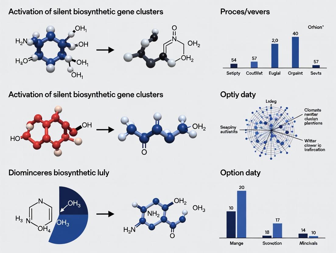
Visualizations
Diagram 2: CRISPR-dCas9 Activation Workflow for a Silent BGC
The Scientist's Toolkit: Research Reagent Solutions
| Item | Function in Silent BGC Research | Example/Supplier |
|---|---|---|
| pCRISPomyces-2 Plasmid | CRISPR-Cas9 system for genetic manipulation (knockout, activation) in Streptomyces. | Addgene #61737 |
| Super Optimal Broth (SOB) Medium | High-efficiency growth medium for E. coli conjugation donors used in intergeneric mating. | Common lab formulation. |
| ERMATE (Ethyl Rhodanine-3-Carboxylic Acid) | Chemical elicitor that mimics competition stress, activates silent BGCs in fungi. | Sigma-Aldrich / Custom synthesis. |
| SACE_5599 (Global Regulator) | Constitutively active mutant of a pleiotropic regulator to boost secondary metabolism in Streptomyces. | Heterologously expressed. |
| ITS1/ITS4 Primers | For rapid fungal DNA barcoding to identify isolates in co-culture studies. | Universal fungal primers. |
| Autoinduction Media (ZYM-5052) | For heterologous expression in E. coli; allows high-density growth before protein production. | Common lab formulation. |
| GNPS (Global Natural Products Social) Platform | Web-based mass spectrometry ecosystem for molecular networking and novelty analysis. | gnps.ucsd.edu |
| antiSMASH Software | In-depth genome mining for BGC prediction and analysis. | antisash.secondarymetabolites.org |
Technical Support Center: Activating Silent Biosynthetic Gene Clusters (BGCs)
Troubleshooting Guides & FAQs
FAQ 1: No Product Detected After Induction of Putative BGC
- Q: I have used a heterologous expression host (e.g., S. albus) and a strong promoter to induce a silent gene cluster, but LC-MS shows no novel metabolites. What are the primary causes?
- A: This is a common challenge. The issue often lies in metabolic reality not matching genomic promise. Refer to the troubleshooting table below.
FAQ 2: Poor Titer of Target Natural Product in Engineered Strain
- Q: My activation strategy worked, but the yield is extremely low for practical purification and characterization. How can I improve titers?
- A: Low titers indicate bottlenecks in the metabolic pathway. Focus on precursor supply, co-factor availability, and alleviating potential toxicity. See the protocols and reagent table.
FAQ 3: How do I choose between in situ activation and heterologous expression?
- Q: For a newly identified silent BGC, what criteria should I use to decide whether to activate it in the native host or clone it into a heterologous host?
- A: The decision matrix below summarizes key factors.
Data Presentation Tables
Table 1: Troubleshooting "No Product" Scenarios
| Potential Cause | Diagnostic Experiment | Possible Solution |
|---|---|---|
| Incorrect Cluster Boundaries | RNA-seq to verify all essential genes are co-transcribed under induction. | Use bioinformatics tools (e.g., antiSMASH deep) to re-predict boundaries; clone extended region. |
| Lack of Pathway-Specific Regulator | Co-express with candidate SARP/LuxR-family regulators from similar clusters. | Clone and co-express a pathway-specific activator gene. |
| Missing Precursor Building Blocks | Supplement growth media with suspected precursors (e.g., amino acids, acyl-CoA). | Engineer host to overproduce key precursors (e.g., malonyl-CoA, methylmalonyl-CoA). |
| Toxic Intermediate or Product | Measure growth curve post-induction; use export gene screening. | Co-express putative resistance/transporter genes from the cluster. |
| Silenced Chromatin State | Perform ChIP-seq for histone marks (if applicable). | Add epigenetic modifiers (e.g., SAHA, 5-azacytidine) to culture. |
Table 2: Heterologous Host Selection Matrix
| Host Strain | Optimal for BGC Type | Key Advantage | Common Limitation |
|---|---|---|---|
| Streptomyces albus J1074 | Type I & II PKS, NRPS | Clean secondary metabolome, high transformation efficiency. | May lack specific tailoring enzymes or co-factors. |
| Pseudomonas putida KT2440 | NRPS, non-ribosomal peptides | Robust growth, solvent tolerance, genetic tools. | Limited experience with complex polyketides. |
| Escherichia coli BAP1 | Type III PKS, simple metabolites | Extremely fast growth, unparalleled molecular tools. | Lacks natural post-translational modifications for many megaenzymes. |
| Mycobacterium smegmatis | Mycobacterial/Actinobacterial clusters | Compatible with unusual lipid precursors. | Slower growth, more complex handling. |
Experimental Protocols
Protocol 1: One-Pot TAR (Transformation-Associated Recombination) Cloning for BGC Capture
- Objective: Capture large (>50 kb) silent BGCs from genomic DNA directly into a yeast-based vector for heterologous expression.
- Methodology:
- Design linear vector and targeting primers with 40-bp homology arms to the 5' and 3' ends of the target BGC.
- Prepare high-molecular-weight genomic DNA from the source organism.
- Co-transform Saccharomyces cerevisiae (e.g., strain VL6-48N) with the linearized capture vector and the genomic DNA using a standard lithium acetate protocol.
- Select for yeast colonies on appropriate synthetic dropout media.
- Isolate yeast plasmid DNA and transform into E. coli for amplification.
- Verify the construct by PCR and restriction digest across several junctions.
Protocol 2: Co-culture Induction for Activating Silent BGCs In Situ
- Objective: Use interspecies microbial interactions to trigger metabolite production from a silent BGC in the native producer.
- Methodology:
- Culture the target strain (harboring the silent BGC) and the inducer strain (e.g., Streptomyces rochei, Bacillus subtilis) separately for 24-48 hours.
- For confrontation assay, spot cultures on opposite sides of an agar plate and incubate until near contact.
- For mixed fermentation, inoculate the target strain and inducer strain (at a 1:1 cell ratio) into the same liquid culture flask.
- Incubate for an extended period (5-10 days).
- Extract metabolites from agar plugs or broth with ethyl acetate and analyze by LC-HRMS.
- Use MS/MS molecular networking (e.g., via GNPS) to identify novel metabolites compared to mono-culture controls.
Mandatory Visualizations
Title: BGC Activation Strategy Decision Workflow
Title: Signaling Pathway for BGC Activation
The Scientist's Toolkit: Research Reagent Solutions
| Reagent / Material | Function in Silent BGC Activation |
|---|---|
| 5-Azacytidine | DNA methyltransferase inhibitor; used for epigenetic derepression of silenced clusters. |
| Suberoylanilide hydroxamic acid (SAHA) | Histone deacetylase (HDAC) inhibitor; relaxes chromatin to promote transcription. |
| N-Acetylglucosamine | Cell wall precursor; often used as a signaling molecule to trigger antibiotic production in Streptomyces. |
| Autoinducer-2 (AI-2) | Quorum-sensing molecule for interspecies communication; used in co-culture elicitation experiments. |
| Chloramphenicol Acetyltransferase (CAT) Reporter System | Coupled with BGC promoter to quantitatively measure activation strength under different conditions. |
| Gateway or Gibson Assembly Kits | For rapid, seamless cloning of large BGC constructs into expression vectors. |
| Codon-optimized tRNA plasmids | For heterologous expression in hosts like E. coli to overcome rare codon usage in native BGC genes. |
| Amberlite XAD-16 Resin | Hydrophobic resin added to fermentations for in situ capture of produced metabolites, reducing feedback inhibition. |
This technical support center provides troubleshooting guidance for researchers working to activate silent biosynthetic gene clusters (BGCs) for novel natural product discovery.
Frequently Asked Questions (FAQs) & Troubleshooting
Q1: My heterologous expression host (e.g., Streptomyces coelicolor) shows no product after introducing a silent BGC. What are the primary causes? A: Failure can stem from multiple factors:
- Lack of Cluster-Specific Activator: The native regulatory gene may not be part of the cloned region or may itself be silent.
- Incompatible Promoters: Native promoters are not recognized by the host's transcription machinery.
- Incorrect Cloning Strategy: The cluster may be fragmented or lack essential regulatory elements.
- Troubleshooting Steps:
- Verify Cluster Integrity: Re-sequence the construct. Use bioinformatics (e.g., antiSMASH) to confirm all genes are present.
- Co-express a Potential Activator: Search for pathway-specific regulator genes (e.g., SARP, LuxR-family) within or near the BGC and clone them under a strong, constitutive promoter.
- Replace Promoters: Use a synthetic biology approach to replace native promoters with strong, constitutive ones (e.g., ermEp, kasOp) for key biosynthetic genes.
Q2: I am using an "OSMAC" (One Strain Many Compounds) approach but see no new metabolites. How can I optimize cultivation parameters? A: OSMAC success depends on systematic variation. Common ineffective parameters include using only standard rich media.
- Troubleshooting Protocol:
- Systematic Media Variation: Prepare a matrix of media with varying C/N sources (e.g., starch, mannitol, soy, chitin), trace elements, and phosphate levels (low phosphate often induces). See Table 1.
- Co-cultivation: Introduce a "challenger" organism (e.g., another actinomycete or fungus) using a split-plate or conditioned media method.
- Add Chemical Elicitors: Supplement with sub-inhibitory concentrations of antibiotics, heavy metals (e.g., 50 µM CuCl₂), histone deacetylase inhibitors (e.g., 10 µM suberoylanilide hydroxamic acid), or N-acetylglucosamine.
Q3: My chromatin remodeling experiment using epigenetic modifiers yielded inconsistent results. What could be wrong? A: Epigenetic manipulation is concentration- and timing-sensitive.
- Troubleshooting Steps:
- Validate Reagent Activity: Use a control strain with a known silent cluster responsive to, e.g., SAHA (a histone deacetylase inhibitor).
- Optimize Treatment Window: Add reagents at different growth phases (early log, mid-log, stationary). Continuous exposure vs. pulse (e.g., 24h) should be tested.
- Check Permeability: Some Gram-positive bacteria have low permeability. Consider using derivatives (e.g., sodium butyrate) or combining with a mild permeabilizing agent like glycine.
Q4: Ribosome engineering (inducing antibiotic resistance mutations) did not activate my target cluster. Should I abandon this approach? A: Not necessarily. This method is stochastic and strain-dependent.
- Optimization Protocol:
- Increase Mutant Library Size: Plate >10⁹ spores/cells on gradient plates with varying levels of antibiotic (e.g., rifampicin, streptomycin). Isolate at least 50-100 resistant mutants.
- Screen Comprehensively: Use a high-throughput method (e.g., LC-MS metabolomics, reporter assay) to screen all mutants, not just a few.
- Combine Strategies: Use a ribosome-engineered mutant as the host for heterologous expression or OSMAC experiments, as its altered translational fidelity can globally affect regulation.
Key Experimental Protocols
Protocol 1: Systematic OSMAC Cultivation for Induction Screening
- Inoculum Prep: Grow seed culture of the target strain for 48h.
- Media Matrix Setup: Prepare 12 distinct media in 250mL flasks (in triplicate). See Table 1.
- Inoculation & Incubation: Inoculate at 2% v/v. Incubate at appropriate temperature with shaking for 7-14 days.
- Sampling: Extract 1mL culture at days 3, 7, and 14 using equal volumes of ethyl acetate (for organic metabolites) and methanol (for polar metabolites).
- Analysis: Concentrate extracts and analyze by HPLC-MS or TLC with appropriate staining.
Protocol 2: Promoter Replacement via λ-RED Recombineering (for E. coli-Actinomycetal Shuttle Vectors)
- Design: Synthesize a linear DNA fragment containing a strong constitutive promoter (e.g., ermEp*) flanked by 50-bp homology arms matching sequences upstream of the target gene's start codon and downstream of its native promoter.
- Prepare Electrocompetent Cells: Induce the λ-RED genes (gam, bet, exo) in the E. coli host carrying the BAC/BAC library clone.
- Electroporation: Electroporate ~100 ng of the linear fragment into competent cells.
- Recovery & Selection: Recover cells in SOC medium for 2h, then plate on appropriate antibiotic.
- Screening: Verify promoter swap by colony PCR and subsequent sequencing.
Data Presentation
Table 1: OSMAC Media Matrix for Silent BGC Activation
| Media Component | Variation 1 (Rich) | Variation 2 (Minimal) | Variation 3 (Stressed) | Variation 4 (Mimic Native) |
|---|---|---|---|---|
| Carbon Source | Glucose (2%) | Glycerol (2%) | Starch (1%) + Mannitol (1%) | Chitin (0.5%) |
| Nitrogen Source | Yeast Extract (0.5%) | NH₄Cl (0.2%) | Soybean Meal (0.3%) | Nitrate (0.1%) |
| Phosphate Level | High (K₂HPO₄, 1 mM) | Low (K₂HPO₄, 0.1 mM) | Very Low (<0.05 mM) | Variable |
| Trace Elements | Standard | + Zn²⁺ (5 µM) | + Fe²⁺ (100 µM) | - |
| pH | 7.0 | 6.5 | 7.2 | 8.0 |
| Elicitor | None | SAHA (10 µM) | N-Acetylglucosamine (5 mM) | Conditioned Media (10% v/v) |
| Reported Success Rate* | ~15% | ~25% | ~35% | ~40% |
Reported success rates are approximate meta-analysis values from recent literature, indicating the percentage of studied strains showing new metabolic profiles under these conditions.
Visualizations
Title: Strategies for Activating Silent Biosynthetic Gene Clusters
Title: Signaling Pathway Leading to BGC Activation
The Scientist's Toolkit: Key Research Reagent Solutions
| Item | Function & Rationale |
|---|---|
| Histone Deacetylase (HDAC) Inhibitors (e.g., SAHA, Sodium Butyrate) | Loosen chromatin structure in eukaryotes and some bacteria, potentially unlocking transcriptionally silent regions of DNA containing BGCs. |
| DNMT Inhibitors (e.g., 5-Azacytidine) | Inhibit DNA methyltransferases, leading to DNA hypomethylation and potential derepression of silenced genes. Used primarily in fungal studies. |
| Ribosome-Targeting Antibiotics (e.g., Rifampicin, Streptomycin) | Used at sub-inhibitory levels to select for spontaneous bacterial mutants with altered ribosomes, leading to global translational and transcriptional changes that can activate BGCs. |
| N-Acetylglucosamine | A cell wall precursor that can act as a signaling molecule, often repressing primary metabolism and activating secondary metabolism in Streptomyces. |
| Autoinducer Molecules (e.g., AHLs, γ-butyrolactones) | Used in co-culture or supplementation studies to probe and activate quorum-sensing pathways that may regulate silent BGCs. |
| Constitutive Promoter Plasmids (e.g., pIJ10257, ermEp) | Vectors for placing key biosynthetic or regulatory genes under strong, constant expression in heterologous hosts to bypass native regulation. |
| λ-RED Recombineering System | Enables efficient, PCR-based promoter replacement or gene knockouts directly on BACs or cosmids carrying large BGCs, without the need for traditional restriction cloning. |
| Broad-Host-Range Expression Vectors (e.g., pSET152, pACYCDuet-1) | Shuttle vectors for moving and expressing BGCs across different bacterial hosts (E. coli, Streptomyces, Pseudomonas). |
Troubleshooting & FAQs
Q1: In my culture-based induction screening, no BGC activation is observed despite using a library of known chemical inducers. What are the potential causes?
A: This failure is common. Primary culprits are:
- Repressor Dominance: The specific repressor protein regulating your target cluster may not be responsive to your inducer library. It may require a very specific, unknown small molecule or a physical signal (e.g., pH, osmolarity).
- Epigenetic Lock: The chromatin state is tightly condensed (heterochromatic). Chemical inducers alone cannot overcome histone deacetylase (HDAC) or DNA methyltransferase activity.
- Lack of Pathway-Specific Activator: The cluster may require a transcriptional activator that is itself silent under your conditions.
Protocol: Co-treatment with Epigenetic Modifiers
- Split your bacterial culture (e.g., Streptomyces) into several flasks.
- Treat with individual inducers from your library in combination with sub-inhibitory concentrations of epigenetic modifiers:
- 5-Azacytidine (DNA methyltransferase inhibitor): 1-5 µM final concentration.
- Suberoylanilide hydroxamic acid (SAHA, HDAC inhibitor): 10-50 µM.
- Incubate for 96-144 hours, sampling every 24h for RNA extraction and metabolomic analysis (e.g., LC-MS).
- Compare gene expression (RT-qPCR of pathway-specific gene) and metabolite profiles to untreated and singly-treated controls.
Q2: My chromatin immunoprecipitation (ChIP) assay confirms histone deacetylation at my target gene cluster, but treatment with broad-spectrum HDAC inhibitors does not activate it. Why?
A: This indicates a multi-layered repression system. Epigenetic silencing is often reinforced by genetic repressors.
- Troubleshooting Steps:
- Perform Dual Inhibition: Combine HDAC inhibitors (e.g., Trichostatin A at 1µM) with a genetic approach to knock down or disrupt a putative pathway-specific repressor gene (identified via bioinformatics).
- Check Histone Methylation Status: Use ChIP-seq with antibodies for H3K9me3 or H3K27me3. These repressive marks are not reversed by HDAC inhibitors and require specific demethylases.
- Verify RNA Polymerase Accessibility: Use an ATAC-seq protocol on treated vs. untreated cells to confirm if chromatin remodeling has actually occurred.
Q3: I have identified a putative repressor gene upstream of my silent BGC. What is the fastest experimental validation workflow?
A: Use a concurrent knockout/complementation and heterologous expression strategy.
Protocol: Repressor Validation Workflow
- In-frame Deletion: Construct an unmarked, in-frame deletion of the putative repressor gene in the native host using CRISPR-Cas9 or homologous recombination.
- Heterologous Expression: Clone the entire BGC (excluding the repressor gene) into an expression vector (e.g., pSET152, BAC library) and express it in a model host (e.g., S. albus).
- Complementation: Express the repressor gene in trans on a plasmid in both the native knockout mutant and the heterologous host.
- Analysis: Measure BGC transcription (RNA-seq) and metabolite production (HPLC-MS) in all four scenarios (Wild-type, ΔRepressor, Heterologous, Heterologous + Repressor).
Q4: How can I distinguish between the lack of an inducer signal and the presence of a strong repressor?
A: A key experiment is the use of global epigenetic derepression as a diagnostic tool.
Protocol: Diagnostic Epigenetic Derepression
- Treat the wild-type strain with a potent, broad-spectrum epigenetic perturbagen cocktail for 72 hours:
- 5-azacytidine (5µM) + SAHA (25µM) + DZNep (Histone methyltransferase inhibitor, 1µM).
- Perform transcriptomics (RNA-seq).
- Interpretation:
- If the BGC remains silent, it strongly suggests a dominant, specific genetic repressor is active.
- If the BGC is activated, the primary gatekeeper is epigenetic silencing. The lack of inducer may be secondary; the native inducer might now be produced by another activated pathway.
The Scientist's Toolkit: Research Reagent Solutions
| Reagent/Category | Example Products/Strains | Primary Function in Activating Silent BGCs |
|---|---|---|
| Epigenetic Modifiers (Chemical) | Trichostatin A (TSA), Suberoylanilide hydroxamic acid (SAHA), 5-Azacytidine, DZNep | Inhibit histone deacetylases (HDACs), DNA methyltransferases (DNMTs), or histone methyltransferases (HMTs) to loosen chromatin compaction. |
| Genetic Toolkits | CRISPR-Cas9 systems (pCRISPomyces), REDIRECT technology (λ-Red recombinering), MAR4 (Conjugative Strain) | Enable targeted gene knockouts (e.g., of repressors), promoter replacements, or entire cluster capture and heterologous expression. |
| Broad-Host Expression Vectors | pSET152, pMS81, BAC (Bacterial Artificial Chromosome) vectors | Shuttle and maintain large DNA inserts (>100kb) in heterologous hosts for expression studies. |
| Model Heterologous Hosts | Streptomyces albus J1074, Mycobacterium smegmatis mc² 155, Pseudomonas putida KT2440 | Clean genetic backgrounds with minimized native secondary metabolism, optimized for expression of foreign BGCs. |
| Inducer Libraries | N-acetylglucosamine, Rare Earth Elements (e.g., La³⁺), Small Molecule Libraries (e.g., from NCI) | Screen for signals that interfere with repressor function or activate pathway-specific regulators. |
| Analytical Standards | Synthetic analogs of predicted core structures, Labeled precursors (¹³C, ¹⁵N) | Essential for metabolomic profiling (LC-HRMS/MS) to identify and characterize novel metabolites produced upon cluster activation. |
Table 1: Efficacy of Common Epigenetic Modifiers in BGC Activation
| Modifier Class | Target | Typical Working Concentration | % of Strains Showing New Metabolites* | Common Off-target Effects |
|---|---|---|---|---|
| HDAC Inhibitor | Histone Deacetylases | 1 - 50 µM | ~15-30% | Growth retardation, altered morphology |
| DNMT Inhibitor | DNA Methyltransferases | 1 - 10 µM | ~5-15% | Genomic instability, high mutation rate |
| HMT Inhibitor | Histone Methyltransferases | 0.5 - 5 µM | ~10-20% | Pleiotropic transcriptional changes |
| Combination (TSA+5-Aza) | HDACs & DNMTs | 1µM + 5µM | ~35-50% | Severe growth inhibition, synergistic toxicity |
*Data aggregated from recent studies (2020-2023) on actinomycete libraries. Efficacy varies greatly by taxonomic group.
Table 2: Success Rates of Common BGC Activation Strategies
| Experimental Strategy | Avg. Time to Result (weeks) | Technical Difficulty (1-5) | Activation Rate (BGC-specific)* | Key Limitation |
|---|---|---|---|---|
| Culture Condition Optimization | 8-12 | 2 | <5% | Highly empirical, low throughput |
| Chemical Epigenetic Perturbation | 2-4 | 1 | 15-30% | Global effects, toxic, not genetic |
| Pathway-Specific Regulator Overexpression | 6-8 | 3 | 20-40% | Requires prior knowledge of regulator |
| Repressor Deletion (CRISPR-Cas9) | 4-6 | 4 | >60% | Requires bioinformatics & genetic system |
| Heterologous Expression | 12-24 | 5 | ~70% | Time-consuming, cloning challenges, possible lack of host factors |
*Defined as detectable transcription of core biosynthetic genes and/or production of a unique metabolite.
Experimental Protocols
Protocol 1: High-Throughput Screening with Epigenetic Elicitors Objective: To rapidly identify strains harboring silent BGCs susceptible to epigenetic derepression.
- Prepare Library: Array bacterial strains in 96-well deep-well plates with 1ml of production medium.
- Treatment: Add epigenetic modifiers (e.g., 5µL of 200x stocks to achieve final concentrations: TSA 1µM, 5-Aza 5µM). Include DMSO-only control wells.
- Incubation: Incubate with shaking (220 rpm) at appropriate temperature for 5-7 days.
- Metabolite Extraction: Add 1ml of ethyl acetate:methanol (3:1) to each well, vortex for 10 min, centrifuge.
- Analysis: Transfer organic layer for LC-MS analysis. Use automated data processing (e.g., MZmine) to detect features unique to treated wells.
Protocol 2: ChIP-seq for Histone Modification Analysis at Silent BGCs Objective: Map repressive histone marks (H3K9me3, H3K27me3) around a silent gene cluster.
- Cross-linking: Grow culture +/- epigenetic treatment. Fix cells with 1% formaldehyde for 15 min. Quench with 125mM glycine.
- Sonication: Lyse cells, sonicate to shear chromatin to 200-500 bp fragments. Confirm size by agarose gel.
- Immunoprecipitation: Incubate chromatin with antibody against target histone mark (e.g., anti-H3K9me3) bound to protein A/G magnetic beads overnight at 4°C.
- Wash, Reverse Cross-link, and Purify: Wash beads stringently, elute DNA, reverse cross-links, and treat with Proteinase K/RNase A. Purify DNA (ChIP-DNA).
- Library Prep & Sequencing: Prepare sequencing library from ChIP-DNA and input DNA (control). Sequence on an Illumina platform (≥5M reads/sample).
- Bioinformatics: Map reads to reference genome, call peaks (e.g., using MACS2). Visualize enrichment over the BGC locus.
Visualizations
Diagram Title: Decision Tree for Silent BGC Activation Failure
Diagram Title: Repressor Validation Experimental Workflow
Diagram Title: Genetic vs Epigenetic Silencing Pathways
Technical Support Center: Troubleshooting Silent BGC Activation Experiments
Frequently Asked Questions (FAQs)
Q1: My heterologous expression host (e.g., S. albus) is not producing the expected compound after BGC insertion. What are the primary causes? A: This is a multi-factorial issue. Common causes include: incorrect promoter recognition in the heterologous host, lack of essential precursor molecules, improper post-translational modification of enzymes, or toxic effects of intermediate compounds. First, verify BGC integrity via sequencing. Then, test different fermentation media and consider co-expressing potential pathway-specific regulator genes from the native host.
Q2: I observe no change in metabolite profile after applying a chemical elicitor (e.g., suberoylanilide hydroxamic acid/SAHA) to my actinomycete strain. How can I troubleshoot? A: 1. Confirm Elicitor Activity: Test the SAHA batch on a control strain (e.g., Streptomyces coelicolor) known to upregulate silent clusters. 2. Optimize Protocol: Ensure you are using an effective concentration (typically 50-100 µM) and adding it at the correct growth phase (often early-mid exponential). 3. Check Detection Sensitivity: Your analytical method (e.g., LC-MS) may not be sensitive enough. Concentrate your culture extract or use longer fermentation times post-elicitation.
Q3: CRISPR-Cas9 activation is not increasing transcription of my target silent gene cluster. What steps should I take? A: 1. Verify gRNA Design: Ensure the gRNA targets the non-template strand near the transcription start site of the putative pathway-specific regulator or cluster boundary. 2. Check dCas9-fusion Protein Expression: Confirm expression of the transcriptional activation domain (e.g., VP64, SoxS) fused to dCas9 via Western blot. 3. Chromatin State: The target region may be in a highly condensed heterochromatin state. Consider combining with histone deacetylase (HDAC) inhibitor treatment.
Q4: My co-culture experiment yields inconsistent results. How can I improve reproducibility? A: Inconsistency often stems from variability in initial cell ratios and environmental conditions. Implement a standardized, reproducible setup:
- Use defined media for both strains.
- Precisely control the starting inoculum ratio (e.g., 1:1, 10:1) using OD600 measurements.
- Consider physical separation methods (e.g., dialysis membranes, partitioned plates) to exchange only diffusible signals, reducing direct competitive growth effects.
Q5: After isolating a novel compound, my antimicrobial assay shows high cytotoxicity against mammalian cell lines but no antibiotic activity. Is this common? A: Yes, this is a known phenomenon in silent BGC activation. Many secondary metabolites evolved as cytotoxins or signaling molecules, not as antibiotics. This compound may have anticancer potential. Proceed with full cytotoxicity profiling against a panel of cancer and non-cancerous cell lines to determine selectivity.
Troubleshooting Guides
Issue: Low Titer of Target Compound in Fermentation
| Potential Cause | Diagnostic Test | Solution |
|---|---|---|
| Suboptimal Growth Conditions | Test growth & production in 4-5 different standard media (e.g., ISP2, R5, SFM). | Switch to the highest yielding medium; perform medium component optimization (e.g., C/N ratio). |
| Inefficient Precursor Supply | Analyze metabolomics data for precursor pool levels (e.g., acetyl-CoA, malonyl-CoA). | Overexpress precursor biosynthetic genes or add benign precursor supplements (e.g., sodium propionate). |
| Inadequate Cluster Induction | qRT-PCR of key biosynthetic genes over time. | Adjust the timing and concentration of chemical inducers; engineer constitutive promoters for pathway regulators. |
Issue: Failed BGC Cloning and Assembly
| Step | Common Problem | Resolution |
|---|---|---|
| DNA Isolation | Sheared or degraded DNA from source strain. | Use gentle lysis protocols; embed mycelia in low-melt agarose plugs for DNA extraction. |
| Capture (e.g., TAR) | No transformants in yeast. | Increase DNA fragment size homology arms to >500 bp; verify yeast recombination strain genotype (rad52). |
| Heterologous Transfer | Vector fails to conjugate into Streptomyces. | Ensure the use of a non-methylating E. coli donor strain (e.g., ET12567/pUZ8002); optimize conjugation conditions (spore age, heat-shock temperature). |
Detailed Experimental Protocols
Protocol 1: One-Strain-Many-Compounds (OSMAC) Screening for BGC Activation Objective: To elicit production of diverse metabolites from a single microbial strain by varying cultivation parameters. Methodology:
- Strain Preparation: Inoculate the strain of interest (e.g., a rare actinomycete) from a glycerol stock onto an agar plate. Grow until sporulation/confluent growth.
- Media Variation: Prepare 10-12 different liquid media in 50 mL flasks. Examples: ISP2, YEME, A3, R2A, GYM, supplemented with variations in carbon source (glucose, maltose, chitin), nitrogen source, or trace elements.
- Inoculation & Cultivation: Inoculate each medium with equal biomass (e.g., 2 mm agar plug). Incubate at appropriate temperature (28°C for actinomycetes) with shaking (220 rpm) for 7-14 days.
- Extraction: Separate biomass and broth by centrifugation. Extract the broth with equal volume of ethyl acetate (x2). Extract the biomass with 1:1 acetone:methanol (x2). Combine organic phases, dry in vacuo.
- Analysis: Redissolve extracts in methanol. Analyze by LC-HRMS (e.g., C18 column, gradient 5-95% acetonitrile in water with 0.1% formic acid, positive/negative ESI). Use metabolomics software (MZmine, XCMS) to compare chromatograms.
Protocol 2: CRISPR-dCas9 Activation of a Target Silent BGC Objective: To specifically upregulate transcription of a chosen silent gene cluster in its native host. Methodology:
- Target Selection & gRNA Design: Identify the putative promoter region of the pathway-specific regulator gene within the BGC. Design two 20-nt gRNA sequences using software (e.g., CHOPCHOP). Clone them into a Streptomyces CRISPR-dCas9 expression plasmid (e.g., pCRISPomyces-2 with VP64 domain).
- Plasmids & Transformation: Verify plasmid by sequencing. Transform into the methylation-deficient E. coli ET12567/pUZ8002 for conjugation.
- Conjugal Transfer: Prepare spore suspension of the target Streptomyces strain. Mix spores with the E. coli donor, plate on MS agar with 10 mM MgCl2. After 16-20h incubation at 30°C, overlay with apramycin (for selection) and nalidixic acid (to counter E. coli).
- Exconjugant Screening: Pick apramycin-resistant exconjugants after 5-7 days. Genotypically confirm via colony PCR for the integrated plasmid.
- Fermentation & Analysis: Cultivate the engineered strain alongside the wild-type in suitable production medium. Harvest at multiple time points. Analyze by qRT-PCR (for transcriptional activation) and LC-MS (for metabolite production).
Diagrams
Title: Experimental Workflow for Silent BGC Activation
Title: Key Signaling Pathways in Microbial Co-culture
The Scientist's Toolkit: Research Reagent Solutions
| Item | Supplier Examples | Function in Silent BGC Research |
|---|---|---|
| HDAC Inhibitors (SAHA, Sodium Butyrate) | Sigma-Aldrich, Cayman Chemical | Chemical epigenetic modifiers that relax chromatin structure, potentially derepressing silent gene clusters. |
| N-Acetylglucosamine | Thermo Fisher, Alfa Aesar | A signaling molecule and component of chitin that can trigger antibiotic production in Streptomyces by altering the activity of global regulators. |
| Autoinducer-2 (AI-2) | Omm Scientific | A universal quorum-sensing molecule used in co-culture studies to mimic bacterial cross-talk and induce secondary metabolism. |
| D-Ala-D-Ala Dipeptide | Bachem, MedChemExpress | A cell wall precursor whose exogenous addition can bypass feedback inhibition, promoting peptidoglycan synthesis and linked antibiotic production. |
| γ-Butyrolactones (e.g., A-Factor) | Custom Synthesis (e.g., AKos) | Streptomycete-specific hormonal signaling molecules that bind receptor proteins to initiate cascade for antibiotic production and morphological differentiation. |
| CRISPomyces-2 Plasmid Kit | Addgene (plasmid #101059) | A modular CRISPR-dCas9 system optimized for Streptomyces for targeted gene activation or repression. |
| Transposon Mutagenesis Kit (EZ-Tn5) | Lucigen | For random insertion mutagenesis to create mutant libraries for discovery of regulatory genes controlling BGC silencing. |
Modern Activation Strategies: From One-Strain-Many-Compounds (OSMAC) to Synthetic Biology
Troubleshooting Guides & FAQs
This technical support center addresses common experimental challenges encountered when applying the OSMAC (One Strain Many Compounds) principle to activate silent biosynthetic gene clusters (BGCs) for natural product discovery.
FAQ 1: After testing multiple media, my microbial strain shows no new metabolite production. What are the primary troubleshooting steps?
- Answer: A lack of observable chemical variation is common. Follow this systematic checklist:
- Analytical Sensitivity: Confirm your detection method (e.g., LC-MS) is sufficiently sensitive and covers a broad metabolite polarity range. The issue may be analytical, not biological.
- Cultivation Time: Harvest cultures at multiple time points (e.g., days 3, 5, 7, 10, 14). Silent clusters may be activated only in late stationary or death phase.
- Sub-culturing: Perform a second passage (sub-culture) of the organism in the promising new medium. Activation sometimes requires physiological adaptation over several generations.
- Biological Replicates: Ensure you have adequate biological replicates (n≥3) to account for natural stochasticity in gene expression.
- Genetic Potential Verification: Re-confirm via genome mining that your strain possesses silent BGCs with high biosynthetic potential.
FAQ 2: How do I choose which OSMAC parameters to vary first for a newly isolated, unsequenced strain?
- Answer: Prioritize parameters based on historical impact and experimental practicality. Follow the tiered approach below.
Table 1: Tiered Prioritization of Initial OSMAC Parameters
| Tier | Parameter | Reason for Priority | Recommended Variations (Initial Screen) |
|---|---|---|---|
| 1 | Culture Medium | Largest impact on primary metabolism and nutrient sensing. | 2-3 distinct complex media (e.g., ISP2, A3M, R5A) and 1-2 defined media. |
| 2 | Aeration/Agitation | Drastically affects oxygen-sensitive regulators and shear stress. | Static, shaken (150 rpm), and highly agitated (250 rpm) conditions. |
| 3 | Ion Concentration | Divalent cations (Fe²⁺, Zn²⁺, Mg²⁺) are common co-factors or signalers. | Add 50-200 µM supplements of FeSO₄, ZnCl₂, MgCl₂, or MnCl₂. |
| 4 | Co-culture | Inter-microbial signaling is a potent activator but adds complexity. | Pair with a phylogenetically distant, non-pathogenic strain (e.g., S. cerevisiae or another Actinobacteria). |
FAQ 3: My LC-MS data shows many new peaks, but dereplication suggests they are known compounds. How can I prioritize novel chemistry?
- Answer: Integrate rapid genomic data with your analytical workflow.
- Perform a quick, low-pass whole-genome sequencing of your strain.
- Use automated tools (antiSMASH, MiBiG) to identify BGCs and predict their core scaffold (e.g., non-ribosomal peptide, polyketide, terpene).
- Correlate MS/MS fragmentation patterns and UV-Vis spectra of your new peaks with the predicted BGC product classes. Peaks whose properties align with a silent (non-expressed under reference conditions) BGC's predicted class are high-priority targets for isolation and structure elucidation.
FAQ 4: What is a robust, standardized protocol for a basic OSMAC screen on actinomycetes?
- Answer: Standardized First-Pass OSMAC Protocol for Actinomycetes
Objective: To induce variation in secondary metabolite profiles of a bacterial isolate.
Materials: See "Research Reagent Solutions" below.
Procedure:
- Inoculum Prep: From a fresh glycerol stock, streak the strain on a standard medium (e.g., ISP2 agar). Incubate until sporulation/confluent growth.
- Seed Culture: Inoculate 50 mL of seed medium (e.g., TSB) in a 250 mL baffled flask with biomass. Incubate at 30°C, 200 rpm for 48h.
- Experimental Variation: Prepare 250 mL baffled flasks each containing 50 mL of different production media (e.g., A3M, R5A, SM14, defined medium with XAD-16 resin).
- Inoculation: Inoculate each production flask with seed culture to a standardized optical density (OD₆₀₀ ≈ 0.1).
- Cultivation: Incubate all flasks at the same temperature (e.g., 30°C) but under two agitation conditions: 220 rpm (high aeration) and 90 rpm (low aeration).
- Harvesting: Collect whole broth samples (cells + supernatant) at 96h, 168h, and 240h.
- Extraction: For each sample, separate supernatant and biomass via centrifugation. Extract supernatant with equal volume of EtOAc. Extract biomass with 1:1 MeOH:DCM. Combine organic extracts, dry in vacuo, and re-dissolve in MeOH for LC-HRMS/MS analysis.
The Scientist's Toolkit: Research Reagent Solutions
Table 2: Essential Materials for OSMAC Experiments
| Item | Function in OSMAC Context | Example/Note |
|---|---|---|
| Baffled Erlenmeyer Flasks | Increases oxygen transfer rate (OTR), a critical variable for aerobic microbes. | 250 mL flask with 50 mL working volume standard. |
| Adsorbent Resins (XAD-16) | Added in situ to adsorb produced metabolites, reducing feedback inhibition and stabilizing labile compounds. | Amberlite XAD-16 resin, autoclaved and added at 1-2% (w/v). |
| Chemically Defined Medium | Allows precise manipulation of individual nutrients (C/N/P source, trace elements) to pinpoint elicitors. | Modify known recipes like Modified R2A or Nitrate Defined Medium. |
| LC-HRMS/MS System | High-resolution mass spectrometry is essential for detecting subtle chemical changes and dereplication. | Q-TOF or Orbitrap systems coupled to UHPLC. |
| Genomic DNA Extraction Kit | For rapid genome sequencing to identify silent BGCs prior to or in parallel with OSMAC screening. | Kits suitable for high-GC content actinomycete DNA. |
| Mining Software (antiSMASH) | Predicts BGCs from genomic data, guiding target prioritization from LC-MS data. | Use web server or local installation (v7+). |
Experimental Workflow & Signaling Pathways
Diagram 1: Tiered OSMAC Experimental Workflow
Diagram 2: Signaling Pathways Linking OSMAC Parameters to BGC Activation
Troubleshooting Guides & FAQs
Q1: My co-culture shows no apparent interaction or activation of new metabolites compared to monocultures. What could be wrong? A: This is often due to insufficient physicochemical interaction or nutrient competition. Ensure your setup allows for metabolite exchange. Use a permeable membrane (e.g., 0.4 µm pore size) if performing spatially separated co-culture. Alternatively, directly mix the strains in a shared medium. Check that your growth media does not overly favor one strain, causing it to dominate and suppress the other. A 1:1 initial inoculum ratio is a starting point, but optimization is required.
Q2: How do I distinguish true chemical induction from simple additive effects from two monocultures? A: You must include analytical controls. Perform:
- Monoculture Controls: Cultivate each strain individually under identical conditions.
- Mixed Extraction Control: Physically mix cell pellets/extracts from harvested monocultures after cultivation but before extraction and analysis.
- True Co-culture: Cultivate strains together from the start. Compare the HPLC-MS or metabolomic profiles of all three. A peak present only in the true co-culture sample indicates an induced or transformed metabolite.
Q3: My interacting co-culture becomes overgrown by a fast-growing fungus, drowning out the bacterial partner. How can I manage this? A: Implement temporal staggering of inoculations. Inoculate the faster-growing organism (e.g., the fungus) 24-48 hours after the slower-growing partner (e.g., the actinomycete). This gives the bacterium a competitive head start. Alternatively, use diffusion chambers or agar-based confrontational assays where physical boundaries can initially separate the organisms.
Q4: What are the best analytical methods to track metabolite exchange and induction in real-time? A: Online monitoring is challenging but possible. Use:
- In-situ Solid-Phase Microextraction (SPE) probes inserted into the broth for continuous sampling of volatile/semi-volatile compounds.
- LC-MS with automated sampling from bioreactors.
- Imaging Mass Spectrometry (e.g., MALDI-TOF IMS) on agar-based co-cultures to spatially map metabolite production at the interaction zone.
Q5: How can I identify which specific molecule from partner A is inducing a BGC in partner B? A: This requires fractionation and activity-guided purification.
- Culture the inducing strain (A) alone.
- Fractionate its culture extract (e.g., by HPLC).
- Add each fraction to monocultures of the responder strain (B).
- Monitor for BGC activation (e.g., via reporter assay, RT-qPCR of pathway genes, or metabolite detection).
- Perform structural elucidation (NMR, HR-MS) on the active fraction.
Q6: My co-culture results are not reproducible between replicates. What steps should I check? A: Focus on standardizing biological and environmental variables:
- Strain Preparation: Use cells from the same growth phase (e.g., mid-log phase) for inoculation. Standardize cryo-stock revival protocols.
- Inoculum Precision: Use optical density (OD) and cell counting for precise, quantitative inoculation.
- Environmental Control: Tightly regulate temperature, shaking speed, and light exposure. For solid media, control agar thickness and drying time.
- Randomized Setup: Arrange biological replicates randomly in the incubator to avoid position-based gradients.
Key Experimental Protocols
Protocol 1: Agar-Based Confrontational Assay for Initial Interaction Screening
Purpose: To rapidly screen for interspecies interactions that may activate silent BGCs. Methodology:
- Streak or spot Strain A on one side of a square agar plate (e.g., ISP2, R2A, or a low-nutrient medium).
- 24-72 hours later, inoculate Strain B on the opposite side.
- Incubate until colonies are nearly confluent (~0.5-1 cm apart).
- Excise agar plugs from: a) the confrontation zone, b) the periphery of each monoculture. Extract separately with ethyl acetate:methanol (3:1).
- Analyze extracts via TLC or HPLC-MS for unique metabolites at the interaction zone.
Protocol 2: Membrane-Separated Co-culture in Liquid Media
Purpose: To allow only chemical exchange while preventing physical contact and cross-predation. Methodology:
- Use a two-compartment vessel (e.g., a cell culture insert with a polycarbonate membrane, 0.4 µm pore size).
- Inoculate Strain A in the main compartment with appropriate medium.
- Inoculate Strain B in the insert compartment.
- Incubate with shaking. Sample compartments independently over time for transcriptomic (RNA-seq) or metabolomic analysis.
- Critical Control: Include "sham" co-cultures where the insert contains sterile medium.
Data Presentation
Table 1: Common Co-culture Systems and Their Applications in BGC Activation
| System Type | Physical Contact? | Key Advantage | Main Disadvantage | Typical BGC Yield Increase* |
|---|---|---|---|---|
| Mixed Liquid | Yes | Maximum interaction potential | Difficult to deconvolve signals | 2x - 10x |
| Membrane-Separated | No | Isolates chemical induction | May miss contact-dependent cues | 1.5x - 5x |
| Agar-Based | Optional (Zonation) | Spatial metabolite mapping | Less scalable for product isolation | 3x - 15x |
| Microfluidic Droplets | Yes/No | High-throughput, single-cell level | Technically demanding | Data pending |
*Reported fold-increase in detectable metabolite classes compared to monoculture averages. Ranges are derived from recent literature surveys (2020-2023).
Table 2: Troubleshooting Common Co-culture Problems
| Problem | Possible Cause | Solution |
|---|---|---|
| No Induction | Lack of stress/nutrient competition | Use low-nutrient media (e.g., M9, PBS with 0.1% carbon source) |
| One strain dominates | Imbalanced growth rates | Stagger inoculation; adjust inoculum ratio; use condition-specific media |
| High replicate variance | Inconsistent inoculation | Standardize to cell count, not OD; use single colony-derived precultures |
| Unclear inducer | Complex metabolite soup | Use fractionated extracts from inducer strain in monoculture assays |
Mandatory Visualizations
Title: Co-culture Workflow for BGC Activation Discovery
Title: Microbial Interaction Pathways Leading to BGC Activation
The Scientist's Toolkit: Research Reagent Solutions
Table 3: Essential Materials for Co-culture Experiments
| Item | Function/Benefit | Example/Specification |
|---|---|---|
| Low-Nutrient Media | Induces stress and competition, mimicking soil/ environmental conditions. | M9 Minimal Media, 10% TSB, PBS with trace carbon. |
| Permeable Membranes | Allows chemical exchange while preventing physical contact. | Polycarbonate inserts, 0.4 µm pore, for 6-well plates. |
| Inactivation/ Fixation Reagents | Snap-freezes microbial community state for 'omics analysis. | RNAprotect for transcriptomics; methanol for metabolomics. |
| Solid Sorbent Probes | For in-situ capture of volatile inducing molecules. | Polydimethylsiloxane (PDMS) coated rods or SPME fibers. |
| Reporter Strains | Visual/quantitative detection of BGC activation. | Strains with GFP fused to promoter of target BGC. |
| Quorum Sensing Inhibitors | Tool to probe role of cell-density signaling. | Synthetic AHL analogs (e.g., furanones), halogenated furanones. |
| Dialysis Culture Apparatus | Scalable chemical exchange co-culture. | Bench-scale dialysis fermentation vessels with 1 kDa membranes. |
FAQs & Troubleshooting Guide
Q1: My heterologous host fails to grow after transformation with the large BGC vector. What could be wrong? A: This is a common issue. Primary causes include:
- Toxic Gene Expression: Unregulated expression of a cluster gene is poisoning the host. Solution: Use a tightly repressible promoter system (e.g., pET with LacI, PBAD with AraC) for initial transformation and growth.
- Vector Burden: The large plasmid size imposes a significant metabolic burden. Solution: Grow cultures at a lower temperature (e.g., 25-30°C) with rich media (e.g., 2xYT, Terrific Broth) and ensure optimal antibiotic concentration—avoid excess.
- Incompatible Replication Origin: The origin's copy number may be unsustainable for large DNA. Solution: Switch to a low or single-copy origin (e.g., SC101, F-derived) for large clusters (>50 kb).
Q2: I have confirmed expression of my BGC in the heterologous host, but no final product is detected. Where should I troubleshoot? A: The issue likely lies in pathway incompatibility or missing precursors.
- Check Precursor Supply: The host may lack essential primary metabolic precursors (e.g., malonyl-CoA, methylmalonyl-CoA for polyketides). Solution: Co-express precursor biosynthetic genes or engineer host pathways to overproduce them.
- Post-Translational Modifications: Your host might lack necessary chaperones or modifying enzymes (e.g., phosphopantetheinyl transferases for NRPS/PKS). Solution: Co-express the cognate sfp-type PPTase from the original organism.
- Silent Regulatory Elements: The native cluster may have embedded regulatory elements not present in your construct. Solution: Re-analyze the intergenic regions for potential small RNA genes or riboswitches; consider constructing a promoter-refactored version.
Q3: I detect unexpected shunt products or intermediates, but not the mature compound. What does this indicate? A: This suggests a bottleneck in the biosynthetic pathway.
- Enzyme Incompatibility: A heterologous enzyme may fold incorrectly or have suboptimal kinetics in the new host. Solution: Optimize codon usage for the host, reduce induction temperature to improve folding, or co-express putative chaperones.
- Failed Tailoring Steps: Late-stage tailoring enzymes (oxidases, methyltransferases, glycosyltransferases) may be inactive. Solution: Verify cofactor availability (e.g., Fe²⁺, SAM, NADPH) in your host and supplement media if needed.
Q4: What are the most critical factors for selecting a heterologous host for silent BGC activation? A: The choice is pivotal. Key factors are summarized below:
| Host Organism | Optimal BGC Type | Key Advantage | Primary Limitation |
|---|---|---|---|
| Streptomyces coelicolor | Actinomycete-derived (Type I/II PKS, NRPS) | Native-like cellular machinery & precursors. | Slow growth, complex genetics. |
| Escherichia coli | Streamlined PKS/NRPS, RiPPs | Fast growth, excellent genetic tools. | Lack of common natural product precursors. |
| Pseudomonas putida | NRPS, Non-ribosomal peptides | Robust metabolism, solvent tolerance. | Fewer specialized toolkits. |
| Saccharomyces cerevisiae | Fungal PKS, Terpenes | Eukaryotic protein processing, compartmentalization. | Low transformation efficiency for large DNA. |
| Bacillus subtilis | RiPPs, Lanthipeptides | Efficient secretion, GRAS status. | Restriction systems can hinder DNA uptake. |
Essential Experimental Protocols
Protocol 1: Gibson Assembly for Refactored BGC Construction
This method is standard for assembling large, promoter-replaced gene clusters.
- Design: Break the target BGC into ~10 kb fragments. Replace native promoters with host-specific, orthogonal promoters (e.g., T7, ermEp*) via PCR. Add 20-40 bp homology overlaps between fragments.
- PCR Amplification: Amplify each refactored fragment and the linearized backbone vector using a high-fidelity polymerase.
- Purification: Gel-purify all DNA fragments.
- Gibson Assembly Reaction:
- Mix equimolar amounts of all fragments and vector (total DNA: 0.02-0.5 pmols).
- Add 2x Gibson Assembly Master Mix (commercial or homemade containing T5 exonuclease, Phusion polymerase, and Taq DNA ligase).
- Incubate at 50°C for 60 minutes.
- Transformation: Desalt the reaction and transform into E. coli competent cells for assembly. Isolate plasmid and subsequently transform into the final expression host.
Protocol 2: Two-Tiered Fermentation for Product Detection
Optimized fermentation is crucial for detecting low-titer compounds.
- Seed Culture: Inoculate a single colony into 5 mL of rich seed medium with antibiotic. Grow at 30°C, 220 rpm for 24-48 hours.
- First-Tier Production (Test Tube Scale):
- Transfer 1 mL seed culture into 25 mL of production medium in a 125 mL baffled flask.
- Induce with optimal concentration of inducer (e.g., 0.5 mM IPTG, 2% mannose) at mid-log phase.
- Incubate for 3-7 days at 20-25°C (to reduce protein misfolding).
- Harvest 1 mL sample daily for LC-MS analysis.
- Second-Tier Production (Bioreactor Scale):
- Scale up the positive culture from Tier 1 to a 1 L bioreactor with 500 mL production medium.
- Strictly control dissolved oxygen (>30%), pH (7.0-7.4), and temperature (25°C).
- Use a fed-batch strategy with limiting carbon source (e.g., glycerol) to maintain metabolic activity without causing repression.
Visualizations
Title: Workflow for Heterologous Expression of Silent BGCs
Title: Logic of BGC Activation via Heterologous Expression
The Scientist's Toolkit: Key Research Reagent Solutions
| Reagent / Material | Function in Heterologous Expression | Example / Note |
|---|---|---|
| Broad-Host-Range Vectors | Shuttle vectors for cloning in E. coli and expression in phylogenetically distinct hosts (e.g., Pseudomonas, Streptomyces). | pUCP series, pRSFDuet, pCAP01 |
| Gibson Assembly Master Mix | Enzymatic mix for seamless, one-pot assembly of multiple linear DNA fragments with homologous overlaps. Critical for refactoring. | New England Biolabs (NEB) HiFi, homemade mix. |
| Methylation-Compatible E. coli | E. coli strains that maintain methylation patterns for DNA to be successfully transformed into hosts with restriction systems. | ET12567 (dam-/dem-), GM2929. |
| Conjugative E. coli Strain | Donor strain for transferring large, non-mobilizable plasmids into Actinomycetes via intergeneric conjugation. | E. coli ET12567/pUZ8002. |
| Tunable Promoter Systems | Precisely regulated promoters for controlling gene expression in heterologous hosts to avoid toxicity. | T7-lac, PBAD, Tip, ermEp* |
| Precursor Supplementation | Chemical additives to supplement host metabolism with limiting co-substrates for biosynthesis. | Sodium propionate, DMB (for cobalamin), SAM. |
| Resin for Metabolite Capture | Hydrophobic resin added to fermentation broth to capture non-polar products, enhancing yield and stability. | XAD-16, Diaion HP-20. |
| LC-MS/MS Grade Solvents | High-purity solvents for metabolite extraction and analysis, minimizing background interference in detection. | Optima LC/MS grade (Fisher). |
Promoter Engineering and Transcription Factor Manipulation
This technical support center provides troubleshooting guidance for experiments in promoter engineering and transcription factor (TF) manipulation, specifically within the context of a thesis focused on activating silent biosynthetic gene clusters (BGCs) for novel natural product discovery. The following FAQs and guides address common experimental pitfalls.
Frequently Asked Questions & Troubleshooting Guides
Q1: My engineered promoter is showing no activity in the heterologous host. What are the primary causes? A: This is often due to host-TF incompatibility. The engineered promoter may lack recognition sites for the host's endogenous RNA polymerase or required transcription factors. Troubleshooting Steps:
- Verify promoter sequence integrity via sequencing.
- Use a reporter gene (e.g., GFP, lacZ) under the control of your promoter in the target host to confirm basal activity.
- Co-express the native or a compatible heterologous TF expected to activate the promoter.
- Check for host-specific silencing mechanisms (e.g., methylation).
Q2: I am attempting to overexpress a pathway-specific transcription factor to activate a silent BGC, but I see no product formation. Why? A: Overexpression alone may be insufficient due to:
- Post-translational Modifications: The TF may require phosphorylation, ligand-binding, or interaction with a co-factor.
- Chromatin State: The target BGC may be in a heterochromatic, silenced state. Consider co-expressing chromatin-remodeling enzymes.
- Incorrect TF: The TF may not be the primary activator for the cluster. Re-analyze bioinformatic predictions.
Q3: My transcription factor manipulation (knock-out/overexpression) leads to severe growth defects or cell death. How can I mitigate this? A: Global TFs can regulate essential genes. Mitigation Strategies:
- Use an inducible expression system (e.g., tetracycline-, arabinose-inducible) for controlled TF expression.
- Employ a conditional knock-out (e.g., CRISPRi for repression) instead of a complete deletion.
- Consider using a modified TF (e.g., a dominant-negative version) that modulates only a subset of targets.
Q4: I have activated a silent BGC and detected a novel metabolite, but the yield is extremely low. How can I improve titers? A: Low titers are common in initial activation. Optimization Approaches:
- Promoter Replacement: Substitute the native promoter of the BGC's core biosynthetic genes with a strong, constitutive, or inducible promoter.
- TF Engineering: Mutate the TF to remove potential regulatory domains that cause repression, creating a constitutively active variant.
- Co-cultivation or Epigenetic Perturbation: Use chemical elicitors (e.g., HDAC inhibitors) or microbial co-culture to mimic natural induction conditions.
Q5: How do I choose between CRISPR-based and homologous recombination-based methods for TF manipulation? A: The choice depends on your host organism and precision needs.
| Method | Best For | Key Advantage | Key Limitation | Typical Efficiency |
|---|---|---|---|---|
| Homologous Recombination | Native hosts with efficient DNA repair (e.g., S. coelicolor, E. coli). | Precise, scarless edits; stable genomic integration. | Can be low-efficiency in non-model hosts; requires long homology arms. | 0.1% - 10% (host-dependent) |
| CRISPR-Cas9 (Knock-out) | Rapid gene disruption in a wide range of hosts. | High efficiency, multiplexing possible. | Off-target effects; requires functional NHEJ or HDR in host. | 50% - 90% in optimized systems |
| CRISPRi/a (Repression/Activation) | E. coli, Streptomyces, fungi; fine-tuning gene expression. | Tunable, reversible, no genomic DNA alteration. | Requires sustained expression of dCas9-protein/fusion. | Repression: up to 99.9% (strong promoters) |
Detailed Experimental Protocols
Protocol 1: CRISPR-dCas9 Mediated Transcriptional Activation (CRISPRa) of a Silent BGC
Objective: To activate a silent gene cluster by recruiting activator domains to its native promoter. Materials: Plasmid expressing dCas9-activator fusion (e.g., dCas9-VPR), sgRNA expression plasmid, transformation equipment, appropriate growth media. Method:
- Design two to three sgRNAs targeting the upstream region of the putative promoter of the target BGC's core biosynthetic gene.
- Clone the sgRNA sequences into your expression vector.
- Co-transform the dCas9-activator and sgRNA plasmids into your host strain.
- Plate on selective medium and incubate.
- Screen colonies (e.g., by PCR for upregulated expression of cluster genes or HPLC for metabolites).
- Validate activation via RT-qPCR comparing transcript levels to a control strain with a non-targeting sgRNA.
Protocol 2: Promoter Swap via Lambda Red Recombineering inE. coli
Objective: To replace the native promoter of a BGC with a strong inducible promoter (e.g., P_tet). Materials: *E. coli* strain with Red system (e.g., BW25141/pKD46), linear DNA cassette (P*tet*_-FRT-antibiotic^R^-FRT), PCR reagents, FLP recombinase plasmid (pCP20). Method:
- Amplify the linear cassette with 50-bp homology arms identical to sequences upstream and downstream of the native promoter region.
- Electroporate the purified cassette into the Red-expressing, electrocompetent host.
- Select for colonies on antibiotic plates corresponding to the cassette marker.
- Verify correct integration by colony PCR using external primers.
- Transform pCP20 to remove the antibiotic marker via FLP-mediated recombination, leaving an FRT scar.
- Verify marker loss and promoter swap by PCR and sequencing.
Diagrams
The Scientist's Toolkit: Research Reagent Solutions
| Item | Function & Application in BGC Activation |
|---|---|
| dCas9-VPR Activation Plasmid | Expresses a catalytically dead Cas9 fused to the VPR transcriptional activator (VP64-p65-Rta). Used in CRISPRa experiments to recruit activation machinery to specific promoter regions. |
| pCRISPomyces-2 Plasmid | A CRISPR-Cas9 system specifically optimized for Streptomyces species, enabling targeted gene knock-outs or promoter replacements to activate or upregulate BGCs. |
| pTet Expression Vector | Plasmid containing a strong, tetracycline-inducible promoter (P_tet_). Used for controlled overexpression of pathway-specific TFs or for promoter swap experiments. |
| HDAC Inhibitors (e.g., Suberoylanilide Hydroxamic Acid - SAHA) | Chemical elicitors that promote a more open chromatin state, potentially de-repressing transcriptionally silent gene clusters in fungi and bacteria. |
| Gateway Cloning System | A versatile, high-efficiency recombination-based cloning system used for rapid assembly of multiple genetic parts (promoters, genes, tags) for TF and promoter engineering constructs. |
| Bacterial Artificial Chromosome (BAC) | Vector for cloning and maintaining large (>100 kb) DNA inserts, such as entire silent BGCs, for heterologous expression studies in tractable hosts. |
| FRT-flanked Antibiotic Cassette | A selectable marker (e.g., apramycin resistance) flanked by FRT sites. Used for selection after genomic integration and subsequently removable via FLP recombinase, leaving a minimal scar. |
Technical Support Center
Troubleshooting Guides & FAQs
Q1: After treating my bacterial/fungal culture with SAHA, I observe no change in metabolite production profile. What could be wrong? A: This is a common issue. First, verify the inhibitor's stability and concentration. SAHA (Vorinostat) is often used in the 1-10 µM range for cell cultures, but effective concentrations for microbial BGC activation can vary. Prepare a fresh DMSO stock solution and avoid repeated freeze-thaw cycles. Ensure your culture medium and conditions (pH, aeration) are suitable for the intended secondary metabolism. Run a positive control, like using a known HDACi (e.g., sodium butyrate) on a model organism. Check Table 1 for typical working concentrations.
Q2: My DNMT inhibitor (e.g., 5-Azacytidine) treatment is causing severe growth inhibition or cell death in my microbial strain. How can I mitigate this? A: DNMT inhibitors like 5-Azacytidine are cytotoxic. Titrate the concentration downwards (start from 0.1 µM) and reduce exposure time. Consider a pulse-treatment protocol: expose the culture for 6-12 hours, then wash and resuspend in fresh medium. This can allow for epigenetic modulation without prolonged cytotoxic stress. Always include a vehicle (DMSO) control and monitor cell density (OD600) throughout.
Q3: I am using a combination of SAHA and a DNMT inhibitor, but my HPLC/MS results show high background noise and inconsistent peaks. A: The inhibitors or their metabolites may co-elute with your compounds of interest. Extract samples with an appropriate organic solvent (e.g., ethyl acetate) to partition inhibitors from polar metabolites. Run a blank sample spiked with only the inhibitors through your analysis pipeline to identify their peak signatures. Consider modifying your chromatographic gradient. Ensure you are quenching metabolism effectively at harvest.
Q4: How do I confirm that HDAC inhibition is actually occurring in my system after SAHA treatment? A: You need a functional readout. Perform Western blot analysis for histone acetylation marks (e.g., H3K9ac, H3K27ac) as a direct pharmacodynamic marker. A significant increase should be visible. Alternatively, use RT-qPCR to monitor transcription of genes known to be immediately responsive to HDAC inhibition in your system as a secondary confirmation.
Q5: I suspect my epigenetic treatment is working, but I cannot detect any new compounds. What analytical strategies should I consider? A: Epigenetic triggers often produce low titers of new metabolites. Implement a guided fractionation approach: Use HR-LC-MS and compare treated vs. untreated extracts with metabolomics software (e.g., MZmine, XCMS) to find features unique to or upregulated in treated samples. Employ molecular networking (GNPS) to visualize related metabolite families. Consider heterologous expression of the activated BGC as a final confirmation.
Data Presentation
Table 1: Common Epigenetic Modulators in BGC Activation Research
| Inhibitor Name | Target | Typical Working Concentration (Microbial Cultures) | Key Considerations |
|---|---|---|---|
| Suberoylanilide Hydroxamic Acid (SAHA, Vorinostat) | HDAC (Class I, II) | 1 – 10 µM | Light-sensitive; prepare in DMSO; check stability in culture pH. |
| Trichostatin A (TSA) | HDAC (Class I, II) | 0.5 – 5 µM | Highly potent; cytotoxic at higher doses. |
| 5-Azacytidine (5-Aza) | DNMT | 0.1 – 10 µM | Incorporate into DNA/RNA; highly toxic; use pulse treatment. |
| Decitabine (5-Aza-2'-deoxycytidine) | DNMT | 0.1 – 5 µM | DNA-specific; more stable than 5-Azacytidine. |
| Sodium Butyrate | HDAC (Class I, IIa) | 1 – 10 mM | Short-chain fatty acid; can affect pH; less potent. |
Table 2: Troubleshooting Summary for Common Problems
| Problem | Possible Cause | Solution |
|---|---|---|
| No BGC Activation | Inhibitor inactive, wrong concentration, BGC refractory. | Use fresh stock, titrate concentration, try combination therapy. |
| Excessive Cell Death | Cytotoxicity of inhibitor. | Reduce concentration/duration, use pulse treatment. |
| No Change in Histone Acetylation | SAHA not cell-permeant or degraded. | Validate with Western blot (H3K9ac), use different HDACi. |
| High Analytical Background | Inhibitors interfere with detection. | Modify extraction protocol, identify inhibitor peaks via blanks. |
| Inconsistent Results | Epigenetic heterogeneity in population. | Biological replicates (n>=3), ensure uniform treatment conditions. |
Experimental Protocols
Protocol 1: Standard SAHA Treatment for Fungal BGC Activation
- Stock Solution: Prepare 50 mM SAHA in high-purity DMSO. Aliquot and store at -20°C protected from light.
- Culture Inoculation: Inoculate your fungal strain into appropriate liquid medium (e.g., YES, PDB). Incubate with shaking (e.g., 150 rpm) at optimal temperature.
- Treatment: At early-mid log phase (e.g., 24-48 h post-inoculation), add SAHA stock to achieve a final concentration of 5 µM (e.g., 1 µL per 10 mL culture). For control, add equal volume of DMSO.
- Incubation: Continue incubation for an additional 3-7 days, depending on growth and production kinetics.
- Harvest: Separate mycelia and broth by filtration. Extract metabolites from both fractions separately with ethyl acetate (2x volume). Pool organic layers, dry over anhydrous Na₂SO₄, and evaporate under reduced pressure.
- Analysis: Resuspend dried extract in methanol for LC-MS analysis.
Protocol 2: Histone Acetylation Analysis by Western Blot (Pharmacodynamic Validation)
- Sample Collection: Harvest cells from treated and control cultures by centrifugation (5,000 x g, 10 min, 4°C).
- Histone Extraction: Use a commercial histone extraction kit or acid extraction (e.g., 0.2 M HCl) for 30 min on ice. Neutralize supernatant and quantify protein.
- Electrophoresis: Load 5-10 µg of histone extract per lane on a 15% SDS-PAGE gel.
- Transfer & Blocking: Transfer to PVDF membrane. Block with 5% BSA in TBST for 1 hour.
- Antibody Incubation: Incubate overnight at 4°C with primary antibody (e.g., anti-H3K9ac, 1:1000). Wash and incubate with HRP-conjugated secondary antibody (1:5000) for 1 hour.
- Detection: Use chemiluminescent substrate and image. Re-probe with total H3 antibody as loading control.
The Scientist's Toolkit: Research Reagent Solutions
| Item | Function in Experiment |
|---|---|
| Suberoylanilide Hydroxamic Acid (SAHA) | Pan-HDAC inhibitor; relaxes chromatin to potentially activate silent BGCs. |
| 5-Azacytidine | DNMT inhibitor; causes DNA hypomethylation, potentially derepressing gene clusters. |
| DMSO (Cell Culture Grade) | Vehicle solvent for dissolving hydrophobic epigenetic inhibitors. |
| Histone Extraction Kit | Isolates histones from cells for downstream modification analysis. |
| Anti-acetyl-Histone H3 (Lys9) Antibody | Primary antibody to detect increased histone acetylation, confirming HDACi activity. |
| HRP-conjugated Secondary Antibody | Enables chemiluminescent detection of Western blot signals. |
| Ethyl Acetate (HPLC Grade) | Organic solvent for broad-spectrum metabolite extraction from culture broth. |
| C18 Reverse-Phase LC Column | For separating complex metabolite mixtures prior to mass spectrometry. |
Pathway & Workflow Diagrams
Title: Epigenetic Activation of Silent BGCs
Title: Epigenetic Screening Workflow
Ribosome Engineering and Global Regulatory Mutants
Troubleshooting Guide & FAQs
This support center is designed to assist researchers in employing ribosome engineering and global regulatory mutations as part of a thesis-focused strategy to activate silent biosynthetic gene clusters (BGCs) for novel natural product discovery.
Frequently Asked Questions
Q1: I've introduced an rpsL (K88E) mutation in my Streptomyces model to confer streptomycin resistance, but I am not observing enhanced antibiotic production. What could be wrong? A: The rpsL (K88E) mutation alters the S12 ribosomal protein, which can pleiotropically affect translation and secondary metabolism. First, confirm the mutation by sequencing the rpsL gene. Second, ensure you are using an appropriate concentration of streptomycin for selection and maintenance (typically 5-50 µg/mL, strain-dependent). Third, consider that the effect might be BGC-specific. Combine this with a second-tier approach, such as introducing a rpoB (H437Y) mutation (rifampicin resistance) or deleting a global repressor like nsdA.
Q2: My global regulatory mutant (e.g., ΔbldA) shows poor sporulation and slow growth, hindering my fermentation scale-up. How can I mitigate this? A: This is a common issue. bldA encodes a tRNA for a rare leucine codon (UUA) found in key developmental and regulatory genes. Poor growth is expected. For fermentation:
- Optimize Media: Switch to a rich, complex medium (e.g., TSB, YEME) for pre-culture and biomass generation before shifting to a production medium.
- Co-culture Strategy: Co-culture the mutant with its parental wild-type strain. The wild-type can provide missing developmental signals or nutritional factors.
- Supplementation: Experiment with adding small molecules (e.g, 0.5% casamino acids, 10 mM MgCl₂) that may bypass metabolic bottlenecks.
Q3: After activating a silent BGC via a rpoB mutation, my LC-MS shows many new peaks. How do I prioritize which to isolate? A: This is a "good problem" to have. Use the following prioritization workflow:
- Dereplication: Compare HR-MS/MS data against natural product databases (e.g., GNPS, AntiBase). Discard known compounds.
- Bioactivity-Guided Fractionation: If your thesis goal is drug discovery, employ a relevant bioassay (e.g., antimicrobial, cytotoxic) to guide isolation.
- Comparative Metabolomics: Compare the metabolite profile of the mutant versus the parent and a "complemented" strain (where the mutation is repaired). Peaks present only in the mutant are directly linked to your engineering strategy.
Q4: I am attempting to combine a ribosomal (rpsL) and a RNA polymerase (rpoB) mutation, but I cannot get a double mutant. What selection strategy should I use? A: Sequential selection is required. Use the following protocol:
Protocol: Generating a DoublerpsL/rpoBMutant
- First Mutation: Introduce the rpsL mutation via selection on streptomycin (Str⁹). Isolate a single, stable mutant (M1).
- Conjugation/Transformation: Introduce a genomic library or perform mutagenesis on M1.
- Double Selection: Plate the resulting cells on media containing both streptomycin (at the concentration used for M1) and rifampicin (5-100 µg/mL). RpoB mutants conferring rifampicin resistance (Rif⁹) arise spontaneously at low frequency.
- Verification: Screen colonies for enhanced or altered metabolite production. Confirm both mutations by PCR amplification and Sanger sequencing of the rpsL and rpoB hot-spot regions.
Table 1: Common Ribosome Engineering Mutations and Their Effects in Streptomyces
| Mutation (Gene) | Antibiotic Resistance | Typical Selection Concentration | Common Observed Phenotype in Secondary Metabolism |
|---|---|---|---|
| K88E (rpsL) | Streptomycin | 5 - 50 µg/mL | Enhanced production of actinorhodin, undecylprodigiosin; activation of silent BGCs. |
| H437Y (rpoB) | Rifampicin | 5 - 100 µg/mL | Drastic changes in transcriptome; frequent activation of silent or cryptic BGCs. |
| K43E (rpsL) | Streptomycin | 2 - 20 µg/mL | Similar to K88E, but severity of effects may differ. |
| P93S (rpsL) | Paromomycin | 10 - 50 µg/mL | Altered aminoglycoside sensitivity; can activate different BGC subsets. |
Table 2: Global Regulatory Mutants for BGC Activation
| Mutated/Deleted Gene | Regulatory Function | Expected Phenotype | Complication for Research |
|---|---|---|---|
| bldA | tRNA for UUA codon | Loss of morphological differentiation; activation of certain BGCs. | Poor growth/sporulation, hard to scale. |
| nsdA | Global repressor | Enhanced antibiotic production; derepression of silent BGCs. | May be species-specific. |
| afsR / afsS | Pleiotropic activator | Overproduction of multiple antibiotics. | Complex regulatory network. |
| rrdA (Rok7B7) | Ribosome silencing factor | Ribosome "de-repression"; activation of specific BGCs. | New target, protocols less standardized. |
Experimental Protocol: Rapid Screening for Activated BGCs Using RIBO-Seq
Objective: To identify translationally activated silent BGCs in a ribosomal mutant versus its wild-type parent.
Method:
- Culture: Grow biological triplicates of wild-type and rpsL (K88E) mutant strains in suitable liquid medium to mid-exponential phase.
- Harvest & Lysis: Rapidly harvest cells by filtration and freeze in liquid N₂. Lyse cells using a bead beater in lysis buffer containing cycloheximide (100 µg/mL) to stall ribosomes.
- Ribosome Protection: Treat lysate with RNase I to digest mRNA not protected by ribosomes. Purify protected mRNA fragments using sucrose cushion ultracentrifugation.
- Library Prep & Sequencing: Isolate RNA from the ribosomal fraction. Prepare a sequencing library (size-select ~28-30 nt fragments). Perform deep sequencing (Illumina).
- Bioinformatics: Map reads to the reference genome. Calculate translational efficiency (TE = RIBO-seq reads / RNA-seq reads) for each gene. Identify BGCs with significantly elevated TE in the mutant but low expression in the wild-type.
Visualizations
Title: RIBO-Seq Workflow for Translational Profiling
Title: Thesis Strategy for Silent BGC Activation
The Scientist's Toolkit: Research Reagent Solutions
| Item | Function in Research | Example/Note |
|---|---|---|
| Streptomycin Sulfate | Selective agent for isolating rpsL (S12) mutants. | Use at 5-50 µg/mL in agar plates for Streptomyces. |
| Rifampicin | Selective agent for isolating rpoB (RNAP β-subunit) mutants. | Use at 5-100 µg/mL. Protect from light. |
| Cycloheximide | Eukaryotic translation inhibitor; used in bacterial RIBO-seq to stall ribosomes during lysis. | Add to lysis buffer (100 µg/mL) to preserve ribosomal complexes. |
| Sucrose Cushion (1M) | Used in ultracentrifugation to purify intact 70S ribosomes and protected mRNA fragments. | Critical for clean RIBO-seq sample preparation. |
| RNase I | Degrades single-stranded RNA; used to digest mRNA not protected by the ribosome. | Preferred over micrococcal nuclease for its specificity. |
| HiScribe T7 High Yield RNA Synthesis Kit | For in vitro transcription of tRNA genes to study ribosome binding/modification effects. | Useful for mechanistic follow-up studies. |
| Methylated UDP-Glucose | Precursor feeding to explore glycosylation patterns in new compounds from activated BGCs. | Part of post-activation structural diversification. |
High-Throughput Screening and Automation in Activation Campaigns
Technical Support Center: Troubleshooting Guides & FAQs
Q1: Our automated liquid handler is consistently delivering inaccurate volumes during the inoculation of 96-well plates with bacterial cultures, leading to poor reproducibility in our gene cluster activation assays. What could be the cause and how can we resolve it?
A1: Inaccurate volume delivery is a common issue. Follow this protocol:
- Calibration Check: Perform a gravimetric calibration on all liquid handling channels using the manufacturer's protocol. Use the same type of tip and labware (e.g., deep-well plate) as in your experiment.
- Tip Integrity: Inspect tips for manufacturing defects. Ensure the tip boxes are correctly seated on the deck.
- Liquid Class Optimization: Review and adjust the liquid class parameters (aspirate/dispense speed, delay, air gap) for your specific culture media. Viscous media may require slower speeds.
- Pre-Wet Step: Add a pre-wetting step (aspirating and dispensing a small volume of liquid to condition the tip interior) to your method.
- Regular Maintenance: Adhere to the scheduled maintenance for wash stations, seals, and gaskets.
Q2: When screening our library of elicitors, we observe high background fluorescence or luminescence in our promoter-reporter assays, obscuring true activation signals. How can we mitigate this?
A2: High background often stems from the media or cellular autofluorescence.
- Media Control: Include wells with media + reporter strain + no inducer, and media alone, to establish baseline.
- Compound Autofluorescence: Pre-screen your chemical libraries for fluorescence/luminescence at your assay's wavelengths. Use a Table of Common Interfering Compounds:
| Compound Class | Typical Interference Wavelength (nm) | Suggested Mitigation |
|---|---|---|
| Polyphenols | 400-550 | Use a red-shifted reporter (e.g., DsRed, mCherry). |
| Alkaloids | 300-450 | Implement a wash step post-elicitation. |
| Quinolones | 350-500 | Employ a secondary, non-optical assay (e.g., RT-qPCR). |
| Media Components (e.g., Yeast Extract) | Broad Spectrum | Switch to a chemically defined minimal media if possible. |
- Reporter Choice: Switch to a longer-wavelength fluorescent protein or a non-optical reporter (e.g., chloramphenicol acetyltransferase).
- Assay Protocol: Centrifuge plates and replace spent media with fresh buffer before reading to remove secreted autofluorescent compounds.
Q3: Our HTS campaign yielded several "hits" that activated our target gene cluster in the primary screen, but these fail to validate in secondary, small-scale flask fermentation. What are the likely reasons?
A3: This discrepancy is often due to differences between microtiter plate (MTP) and flask physiology.
- Aeration/Mixing: MTPs have poor, static gas exchange compared to shaking flasks. The hit compound's effect may be oxygen-sensitive.
- Evaporation: Significant evaporation in MTP edges can concentrate compounds and salts, creating false positives.
- Protocol for Validation: Always include a "Matrix Validation" step:
- Repeat the primary screen hit in the MTP format using staggered, staggered inoculation times.
- Use the same culture volume-to-vessel ratio when scaling to flasks.
- Re-supplement the hit compound in flasks at multiple time points to mimic MTP conditions.
- Monitor pH and dissolved oxygen if possible; differences here can drastically alter gene expression.
Q4: During automated image analysis of colony-based assays, the software is incorrectly segmenting overlapping microbial colonies. How can we improve accuracy?
A4: Improve segmentation by preprocessing images and adjusting parameters.
- Image Acquisition: Use high-resolution scanning and ensure even, diffuse lighting to minimize shadows.
- Pre-processing: Apply background subtraction and a mild Gaussian blur to reduce noise before segmentation.
- Algorithm Tuning: If using a circular Hough transform, adjust the minimum and maximum allowed colony diameter. For watershed-based algorithms, carefully tune the threshold for seed detection and the parameters controlling watershed line placement.
- Manual Review & Training: For complex morphologies, use machine learning-based tools. Manually label a subset of images (correct vs. overlapping colonies) to train a classifier for improved recognition.
The Scientist's Toolkit: Research Reagent Solutions
| Item | Function in Activation Campaigns |
|---|---|
| Chemical Elicitor Libraries (e.g., NPL, Prestwick) | Diverse collections of bioactive molecules (HDAC inhibitors, receptor ligands, antibiotics) used to perturb cellular regulation and potentially activate silent BGCs. |
| Broad-Host-Range Reporter Plasmids (pPROBE, pMS) | Plasmid vectors carrying promoterless fluorescent/luminescent genes. They can be integrated into or transferred across diverse bacterial strains to monitor promoter activity of target BGCs. |
| Ribosome Engineering Antibiotics (e.g., Rifampicin, Streptomycin) | Low-dose antibiotics used to generate genetic mutations, particularly in ribosomal proteins, leading to pleiotropic effects that can globally upregulate secondary metabolism. |
| Quorum Sensing Molecules (AHLs, AIPs, γ-butyrolactones) | Used as co-culture supplements or to mimic microbial interactions, tricking bacteria into activating BGCs normally reserved for competitive or cooperative situations. |
| HDAC/DAC Inhibitors (Suberoylanilide Hydroxamic Acid, 5-Azacytidine) | Epigenetic modifiers that alter chromatin structure in fungi or DNA methylation in bacteria, potentially unlocking transcriptionally silent gene clusters. |
| Resin-Assisted Capture Media | XAD resins added to fermentation broths to adsorb secreted natural products, reducing feedback inhibition and increasing titers for detection. |
Experimental Protocol: Primary HTS for Elicitor Discovery
Objective: To identify small molecules that activate a silent biosynthetic gene cluster (BGC) in a high-throughput manner. Method:
- Strain Preparation: Transform your bacterial strain harboring the silent BGC with a plasmid where a key cluster promoter (P~BGC~) drives a GFP reporter gene. Prepare a glycerol stock of the reporter strain.
- Plate Setup: Using an automated liquid handler, dispense 95 µL of appropriate growth medium into each well of sterile 96-well microtiter plates.
- Compound Dispensing: Pin-transfer (nL-µL volumes) or acoustically dispense compounds from the chemical library into assay plates. Include controls: DMSO-only (negative), a known inducer if available (positive).
- Inoculation: Thaw the reporter strain glycerol stock, dilute in medium to an OD~600~ of 0.05. Dispense 5 µL per well using the liquid handler, achieving a final OD~600~ of ~0.0025.
- Incubation: Seal plates with a breathable membrane and incubate at optimal growth temperature with shaking for 24-72 hours.
- Signal Detection: Measure optical density (OD~600~) for growth and fluorescence (Ex/Em ~488/510 nm) for promoter activity using a plate reader.
- Data Analysis: Calculate the activation ratio (Fluorescence/OD~600~) for each well. Normalize to the median of the DMSO controls. Hits are typically defined as compounds causing an activation ratio > 3 standard deviations above the normalized median.
Visualizations
HTS Screening Workflow for BGC Activation
Putative Pathways for Elicitor-Induced BGC Activation
Overcoming Activation Hurdles: Troubleshooting Failed Expression and Yield Optimization
Troubleshooting Guides & FAQs
Q1: After inducing our silent BGC in the heterologous host, we detect no product. What are the primary diagnostic steps? A1: The first step is a two-pronged approach to confirm (1) genetic integrity and (2) transcriptional competence.
- Cluster Integrity Check: Perform long-range PCR or whole-plasmid sequencing across the entire cloned BGC to rule out deletions or mutations during cloning. Compare the sequence to the reference.
- Pathway Expression Check: Use RT-qPCR on key biosynthetic genes (e.g., the core synthase gene) to confirm mRNA is being produced. A lack of product despite confirmed mRNA shifts the focus to translation, enzyme folding, or substrate availability.
Q2: Our RT-qPCR shows the cluster is being transcribed, but the metabolite is not detected. What could be wrong? A2: Transcription without production points to post-transcriptional failures. Key troubleshooting areas include:
- Codon Bias: Heterologous hosts may inefficiently translate genes with rare codons. Check codon adaptation indices (CAI) and consider supplying tRNA plasmids.
- Protein Folding/Activity: The host may lack necessary chaperones or post-translational modifications. Check for required phosphopantetheinyl transferase activity for NRPS/PKS systems.
- Precursor Limitation: The host's native metabolism may not supply sufficient building blocks (e.g., malonyl-CoA, amino acids). Consider precursor feeding or engineering host metabolic pathways.
- Toxic Intermediates: Accumulation of pathway intermediates may be toxic, halting production.
Q3: How can we definitively determine if a silent BGC is intact in the native genome before attempting cloning? A3: Use a combination of bioinformatic and molecular techniques.
- Bioinformatic Analysis: Assemble genomes from high-coverage sequencing. Use tools like antiSMASH to predict BGC boundaries and check for internal stop codons, frameshifts, or transposon insertions.
- PCR Survey: Design overlapping PCR amplicons tiling across the predicted BGC. Successful amplification suggests physical continuity, while failed reactions indicate potential breaks.
Q4: What are common reasons for pathway silence in the native host, and how can we overcome them for expression? A4: Common causes and strategies are summarized below:
| Silence Cause | Mechanism | Diagnostic/Activation Strategy |
|---|---|---|
| Lack of Inducer | Repressor protein bound; no signal molecule. | Use chromatin immunoprecipitation (ChIP) to identify repressors; screen signal molecules (e.g., N-acetylglucosamine). |
| Heterochromatin | Histone deacetylases (HDACs) condense chromatin. | Treat with HDAC inhibitors (e.g., suberoylanilide hydroxamic acid); quantify histone modification marks (H3K9ac). |
| Weak/Non-Functional Promoter | Native promoter not recognized by host machinery. | Replace with a strong, constitutive or inducible promoter (e.g., PtipA, ermE*p). |
| Absence of Pathway-Specific Regulator | Regulator gene is missing, mutated, or located elsewhere. | Clone and co-express putative regulator genes; use CRISPRa to activate native regulator expression. |
Experimental Protocols
Protocol 1: Diagnostic PCR for BGC Integrity Objective: To verify the physical continuity and absence of major deletions in a cloned biosynthetic gene cluster. Materials: High-fidelity DNA polymerase (e.g., Q5), primers flanking ~5-10 kb intervals across the BGC, cloned BGC DNA as template. Procedure:
- Design primer pairs to generate overlapping amplicons that tile across the entire BGC (e.g., 8-12 amplicons for a 50 kb cluster).
- Set up PCR reactions according to the high-fidelity polymerase protocol. Use an extension time appropriate for the amplicon length (1 min/kb).
- Run the PCR products on a 0.8% agarose gel.
- Expected Outcome: All amplicons should produce single bands of expected size. A missing or size-shifted band indicates a deletion or rearrangement at that locus.
Protocol 2: RT-qPCR for Transcriptional Analysis of a Silent BGC Objective: To quantify expression of genes within a potentially silent biosynthetic gene cluster. Materials: RNA extraction kit, DNase I, reverse transcription kit, SYBR Green qPCR master mix, gene-specific primers. Procedure:
- RNA Extraction: Extract total RNA from cultures under test and control conditions. Include a biological replicate.
- DNase Treatment: Treat RNA with DNase I to remove genomic DNA contamination.
- cDNA Synthesis: Perform reverse transcription using random hexamers.
- qPCR Setup: Prepare reactions with SYBR Green master mix, cDNA template, and primers for your target BGC gene(s) and a stable reference gene (e.g., rpoB or hrdB).
- Run & Analyze: Perform qPCR. Use the ΔΔCt method to calculate the fold-change in expression of target genes relative to the reference gene and the control condition.
Visualizations
Title: Primary Diagnostic Workflow for Silent BGC Activation
Title: Transcriptional Activation Pathway of a Silent BGC
The Scientist's Toolkit: Research Reagent Solutions
| Item | Function in Silent BGC Research |
|---|---|
| HDAC Inhibitors (e.g., SAHA) | Loosen condensed chromatin to potentially unlock transcriptionally silent clusters in native hosts. |
| Phosphopantetheinyl Transferase (PPTase) | Essential for activating carrier proteins in NRPS/PKS systems; required when expressing clusters in hosts lacking compatible PPTases. |
| Rare tRNA Supplement Kits | Enhance translation efficiency of heterologously expressed genes with non-optimal codon usage for the host. |
| Inducible Promoter Systems (Ptet, PtipA) | Provide precise, strong control over the expression of the entire cloned BGC in a heterologous host. |
| CRISPR Activation (CRISPRa) System | Used to target and activate transcription of silent BGCs directly in their native genomic context. |
| Stable Isotope-Labeled Precursors (e.g., ¹³C-Acetate) | Feed to cultures to confirm pathway activity and trace metabolite production via LC-MS, even at low yields. |
Optimizing Heterologous Hosts (Streptomyces, Aspergillus, E. coli) and Vector Systems
Technical Support Center: Troubleshooting Guides & FAQs
Frequently Asked Questions (FAQs)
Q1: My silent biosynthetic gene cluster (BGC) shows no expression in E. coli. What are the primary troubleshooting steps? A: First, verify codon optimization for E. coli. Second, check for toxic gene products by inducing with lower inducer concentrations (e.g., 0.1 mM IPTG). Third, ensure the presence of necessary post-translational modification machinery or co-expression of relevant enzymes. Fourth, test alternative promoters (e.g., T7, trc) and ribosome binding sites.
Q2: In Streptomyces, I am getting very low titers of my target compound. How can I improve yield? A: Low titers can be addressed by: 1) Using strong, constitutive promoters (e.g., ermEp, *kasOp) instead of native ones. 2) Optimizing media composition (carbon/nitrogen source). 3) Co-expressing precursor biosynthesis genes. 4) Deleting competing pathway genes or global regulators. 5) Extending fermentation time and sampling.
Q3: My Aspergillus transformation is inefficient, resulting in very few positive transformants. What could be wrong? A: Key factors are protoplast quality and DNA purity. Ensure young, actively growing mycelia are used for protoplast generation. Use fresh lysing enzymes. Purify vector DNA via cesium chloride gradient or equivalent high-purity method. Include an osmotic stabilizer (e.g., 1.2 M sorbitol) in all post-protoplast steps.
Q4: I suspect plasmid instability in my Streptomyces heterologous host over long-term cultivation. How can I confirm and fix this? A: Confirm by performing plasmid rescue from aged cultures and comparing to original. To improve stability: 1) Use integrative vectors (e.g., based on ΦC31, BT1 integrase) for chromosomal insertion. 2) If using a replicative vector, include a selective antibiotic throughout cultivation. 3) Use a host deficient in major restriction systems.
Q5: Protein expression from my BGC in E. coli results in insoluble inclusion bodies. What are my options? A: Strategies include: 1) Lowering expression temperature (e.g., 18-25°C). 2) Reducing inducer concentration. 3) Using a weaker promoter. 4) Co-expressing molecular chaperones (e.g., GroEL/ES). 5) Switching to a fusion tag system (e.g., MBP, GST) known to enhance solubility. 6) Testing different E. coli strains (e.g., Origami for disulfide bonds).
Troubleshooting Guides
Issue: Failed Intergeneric Conjugation from E. coli to Streptomyces.
- Check 1: Donor E. coli strain. Ensure you are using a methylation-deficient E. coli donor (e.g., ET12567/pUZ8002) to bypass Streptomyces restriction barriers.
- Check 2: Conjugation mixture. Use a 1:1 ratio of donor spores to recipient mycelia. Ensure the presence of 10 mM MgSO4 on the conjugation plate.
- Check 3: Antibiotics. Use the correct selective agents for the exconjugants. Overlay antibiotics after 16-24 hours of co-incubation.
Issue: No Heterologous Production in Aspergillus nidulans despite successful transformation.
- Step 1: Verify integration. Perform diagnostic PCR from genomic DNA across integration junctions to confirm correct locus-specific integration.
- Step 2: Check promoter-gene fusion. Re-sequence the cloned construct in the expression vector to ensure the BGC genes are correctly fused to the fungal promoter (e.g., gpdA, alcA) and terminator.
- Step 3: Assess culture conditions. Test different production media (e.g., minimal media vs. complex) and induction conditions. For inducible promoters, verify the inducing agent (e.g., threonine for alcA).
Issue: Poor Vector Yield from E. coli Cloning Stalls for Large BGC Constructs (>30 kb).
- Action 1: Use specialized strains. Clone large cosmids/BACs in high-copy-number E. coli strains like EPI300, which allow inducible copy number amplification.
- Action 2: Optimize DNA isolation. Use alkaline lysis methods scaled for large plasmids, followed by isopropanol precipitation. Consider additional purification via anion-exchange columns.
- Action 3: Verify stability. Perform restriction analysis of the isolated plasmid compared to the original to check for deletions or rearrangements.
Table 1: Comparison of Key Features for Heterologous Host Systems in BGC Activation
| Feature | E. coli | Streptomyces spp. | Aspergillus spp. |
|---|---|---|---|
| Typical BGC Size Limit | < 70 kb (Cosmid/BAC) | > 100 kb (Cosmid/BAC) | 30-80 kb (Fosmid/Cosmid) |
| Transformation Efficiency | 10^7 - 10^9 CFU/µg (plasmid) | 10^4 - 10^6 CFU/µg (plasmid) | 10 - 100 transformants/µg (integrative) |
| Growth Speed | Fast (hours) | Slow (days) | Moderate (days) |
| Native PTM Capability | Low | High (methylation, prenylation) | High (glycosylation, oxidation) |
| Common Vector Type | High-copy plasmids, BACs | Integrating vectors, Shuttle cosmids | Integrating vectors, AMA1-based plasmids |
| Key Advantage | Rapid genetics, high yield proteins | Native-like expression for actinomycete BGCs | Efficient secretion, eukaryotic PTMs |
| Major Challenge | Lack of complex PTMs, toxicity | Slow growth, complex genetics | Lower transformation efficiency, harder genetics |
Table 2: Common Vector Systems and Their Applications
| Vector System | Host Range | Copy Number | Key Application for Silent BGCs |
|---|---|---|---|
| pET Series | E. coli | High (upon induction) | High-level expression of individual BGC enzymes |
| pRSET Series | E. coli | High | Solubility-tagged protein expression for activity assays |
| pIJ86/pSET152 | Streptomyces | Single (integrative) | Stable integration and expression of full BGCs |
| pKC1139 | Streptomyces | Low-copy (replicative) | Shuttle vector for library construction and conjugation |
| pTYGS Series | Streptomyces | Varies | Type III polyketide synthase expression and engineering |
| pTAex3 | Aspergillus | Low (integrative) | Strong gpdA promoter-driven expression in fungi |
| pFC902 | Aspergillus | Autonomous (AMA1-based) | High-copy maintenance for gene overexpression |
Experimental Protocols
Protocol 1: Intergeneric Conjugation for Streptomyces Transformation
- Preparation: Grow E. coli ET12567/pUZ8002 donor strain (carrying the orIT-containing vector) in LB with appropriate antibiotics to mid-log phase.
- Wash: Harvest cells, wash 3x with LB to remove antibiotics.
- Recipient: Prepare Streptomyces spores or mycelia, treat with 50°C heat shock for 10 minutes if using spores.
- Mixing: Mix donor and recipient cells in a 1:1 ratio, pellet, and resuspend in a small volume.
- Plating: Spot the mixture onto MS agar (with 10 mM MgSO₄). Dry and incubate at 30°C for 16-24 hours.
- Selection: Overlay plate with sterile water containing nalidixic acid (to counterselect E. coli) and the antibiotic for plasmid selection. Incubate until exconjugants appear (3-7 days).
Protocol 2: Protoplast Preparation and Transformation for Aspergillus nidulans
- Mycelia Growth: Inoculate 1x10^8 conidia into 100 ml liquid minimal media. Incubate overnight (16-18h) at 37°C, 220 rpm.
- Harvest: Filter mycelia through Miracloth, wash with 0.6 M KCl.
- Digestion: Resuspend mycelia in 20 ml digestion solution (10 mg/ml lysing enzymes, 0.6 M KCl). Incubate 2-3h at 30°C, 80 rpm.
- Protoplast Isolation: Filter through Miracloth, centrifuge filtrate at 2500 g for 10 min. Wash pellet gently with 1.2 M sorbitol, 50 mM CaCl₂ (STC).
- Transformation: Mix 100 µl protoplasts (~10^7), 5-10 µg DNA (in STC), and 50 µl 60% PEG-4000 (in STC). Incubate on ice 20 min, then add 0.5 ml 60% PEG-4000, mix, incubate at room temp 5 min.
- Regeneration: Add STC, plate onto regeneration agar (with 1.2 M sorbitol). After 24h, overlay with selective agar.
Diagrams
Title: Heterologous BGC Activation Workflow
Title: Key Factors for BGC Activation
The Scientist's Toolkit: Research Reagent Solutions
| Reagent/Material | Function in Experiment |
|---|---|
| Cosmid/Fosmid Vectors (e.g., pCC1FOS) | Cloning and stable maintenance of large (>30 kb) BGC DNA fragments in E. coli. |
| Gateway or Gibson Assembly Kits | Seamless cloning and assembly of multiple BGC fragments into expression vectors. |
| E. coli Strain ET12567 | Donor strain for intergeneric conjugation; dam-/dem- to avoid restriction in Streptomyces. |
| Streptomyces Integration Vector pSET152 | ΦC31 attP/int-based vector for stable, single-copy integration of BGCs into the host chromosome. |
| Aspergillus Expression Vector pTAex3 | Contains strong, constitutive gpdA promoter for driving BGC expression in fungal hosts. |
| Lysing Enzymes from Trichoderma harzianum | Digest fungal cell walls to generate protoplasts for Aspergillus transformation. |
| ISP2/MS Agar Media | Standard media for Streptomyces conjugation, sporulation, and secondary metabolism. |
| Terrific Broth (TB) Media | High-density growth medium for E. coli to yield large amounts of plasmid/cosmid DNA. |
| HisTrap HP Column | For rapid purification of His-tagged recombinant proteins expressed from BGC genes in E. coli. |
| C18 Solid-Phase Extraction (SPE) Cartridges | For desalting and concentrating crude culture extracts prior to LC-MS analysis of metabolites. |
Addressing Toxicity of Pathway Intermediates or Final Products
Technical Support Center
Troubleshooting Guides & FAQs
Q1: My heterologous host strain exhibits severe growth inhibition or cell lysis upon induction of a target silent gene cluster. How can I determine if this is due to toxicity from an intermediate or final product? A: This is a classic sign of toxicity. To diagnose the source, implement a tiered approach:
- Metabolite Profiling: Use LC-MS to analyze culture supernatants and cell lysates at multiple time points post-induction. Compare profiles to uninduced controls.
- Genetic Dissection: Systematically inactivate individual genes in the cluster (e.g., via promoter disruption or in-frame deletion) and observe the host growth phenotype upon induction. Restoration of normal growth pinpoints the step producing the toxic compound.
- Intermediate Feeding: If possible, chemically synthesize predicted early pathway intermediates and feed them to cultures. Observed toxicity can map the problem to a specific compound.
Q2: I have identified a toxic intermediate. What strategies can I employ to stabilize my production host? A: Several metabolic engineering and cultivation strategies can mitigate toxicity:
| Strategy | Mechanism | Example/Protocol |
|---|---|---|
| Promoter Engineering | Fine-tune expression of toxic biosynthetic steps. | Use a tunable promoter system (e.g., Ptet, PBAD) to express the problematic gene(s). Perform a gradient induction experiment to find a level that balances production and growth. |
| Export Pump Expression | Actively transport toxic compounds out of the cytoplasm. | Heterologously express known efflux pumps (e.g., Bacillus BmrA, E. coli TolC) or putative transporters from the native cluster host. Clone pump gene(s) into production plasmid. |
| Enzyme Compartmentalization | Secrete enzymes or localize pathway to reduce cytoplasmic toxicity. | Fuse signal peptides (e.g., PelB, OsmY) to pathway enzymes for periplasmic secretion. Alternatively, use synthetic protein scaffolds to create metabolic microcompartments. |
| Two-Stage Fermentation | Decouple growth from production. | Protocol: 1) Grow culture to mid-log phase in optimal growth medium. 2) Harvest cells via centrifugation. 3) Resuspend in production medium (e.g., nitrogen-limited) and induce cluster expression. Monitor viability and titers over 24-72h. |
| In Situ Product Removal | Continuously extract product from culture. | Integrate an adsorbent resin (e.g., XAD-16, HP20) or a second immiscible organic phase (e.g., dioctyl phthalate) into the bioreactor. This sequesters the toxic compound. |
Q3: The final product of my activated cluster is highly cytotoxic, complicating purification and analysis. How can I handle this? A: Modify handling protocols and consider bioengineering solutions.
- FAQs:
- Q: How should I quench my culture for analysis?
- A: Immediately mix culture with an equal volume of cold quenching solvent (e.g., 60% methanol, -40°C) to halt metabolism. Process samples on ice or at 4°C.
- Q: What chromatography considerations are important?
- A: Use inert HPLC systems (e.g., PEEK tubing and fittings) and pre-saturate columns with high concentrations of product to prevent adsorption losses. Consider online dilution for MS injection.
- Q: Can I modify the product to reduce toxicity?
- A: Yes. If the mode of action is non-essential for initial study (e.g., you are characterizing scaffold production), consider mutating the final tailoring enzyme (e.g., an acyltransferase) to produce a less toxic analog.
Experimental Protocol: Rapid Toxicity Assessment via Spot Assay Purpose: To quickly screen multiple engineered strains for improved tolerance to a toxic compound or pathway. Materials:
- Solid agar plates: Base growth medium.
- Top agar: 0.7% agar in growth medium, kept liquid at 48°C.
- Overnight cultures of test strains (e.g., control, efflux pump-expressing, promoter-variant).
- Inducer compound (if testing pathway toxicity). Method:
- Serially dilute overnight cultures (100 to 10-5) in sterile saline.
- Mix 100 µL of each dilution with 3 mL of molten top agar. For induction, add the required inducer to the top agar tube before mixing.
- Pour the mixture immediately onto a base agar plate. Allow to solidify.
- Incubate plates at appropriate temperature for 24-48 hours.
- Interpretation: Compare the highest dilution yielding colony growth between strains. Improved tolerance is indicated by growth at higher dilutions under inducing conditions.
Toxicity Troubleshooting Workflow for Activated BGCs
Research Reagent Solutions
| Item | Function | Example/Supplier |
|---|---|---|
| Tunable Expression Vectors | Enables precise control of gene expression levels to balance production and toxicity. | pET Duet vectors (Novagen), pCOLA Duet (Addgene), T7 RNA polymerase/promoter system with LacI regulation. |
| Broad-Host-Range Cloning Kits | For expressing putative efflux pumps or regulators in diverse heterologous hosts. | CloneEZ Hi-Blunt Kit (GenScript), Gibson Assembly Master Mix (NEB). |
| Adsorbent Resins | For in situ product removal (ISPR) to sequester toxic compounds from culture broth. | Amberlite XAD-16N (Sigma-Aldrich), Diaion HP20 (Supelco). |
| Quenching Solvent | Rapidly stops cellular metabolism for accurate snapshot of intracellular metabolites. | 60% Aqueous Methanol (-40°C). |
| PEEK HPLC Hardware | Inert fluidic path to prevent adsorption of reactive or toxic compounds during analysis. | PEEK tubing, fittings, and injection loops (e.g., Idex Health & Science). |
| Biosynthetic Precursors | Feeding studies to pinpoint toxic steps and potentially shunt metabolism. | Sodium propionate, malonyl-CoA, methylmalonyl-CoA, specialized amino acids (Sigma-Aldrich). |
| Whole-Cell Biosensors | Reporters for real-time detection of stress (e.g., membrane damage, SOS response) upon induction. | Strains with stress-responsive promoters (e.g., PrecA, PkatG) fused to GFP/luciferase. |
This technical support center provides guidance for researchers working on activating silent biosynthetic gene clusters (BGCs) through tiling and refactoring approaches. These methods are central to modern natural product discovery, enabling the simplification and heterologous expression of complex genetic architectures to unlock novel bioactive compounds.
Troubleshooting Guides & FAQs
Q1: During BGC refactoring, my heterologous host fails to produce the expected compound. What are the primary troubleshooting steps? A: This is a common issue. Follow this systematic approach:
- Verify Cluster Integrity: Sequence the entire refactored cluster to confirm no errors were introduced during synthesis/assembly. Pay special attention to promoter and RBS sequences.
- Check Expression Cascade: Use RT-qPCR to analyze transcription of each gene in the refactored operon. A failure in one gene can halt the entire pathway.
- Assess Host Compatibility: Ensure essential precursors (e.g., acyl-CoA units, amino acids) are available in your host (E. coli, Streptomyces, etc.). Consider supplementing the media or engineering host precursor pathways.
- Test Enzyme Functionality: Express and assay key enzymes (e.g., polyketide synthases, non-ribosomal peptide synthetases) in vitro to confirm correct folding and activity in the heterologous context.
Q2: How do I decide between tiling (e.g., CRISPR-Cas9 aided) versus complete de novo refactoring for a large, silent BGC? A: The choice depends on cluster complexity, genetic accessibility, and project goals.
| Factor | Tiling (In situ Editing) | De novo Refactoring |
|---|---|---|
| Best For | Clusters in cultivable, genetically tractable native hosts. | Clusters from uncultivable hosts or extremely complex regulation. |
| Speed | Faster if host tools exist. | Slower due to design, synthesis, and assembly. |
| Control | Limited to native regulatory elements. | Complete control over promoters, RBSs, and gene order. |
| Primary Risk | Off-target edits; native host physiology may still silence production. | Incorrect assembly; synthesized genes may have functional errors. |
| Protocol Reference | See Protocol 1: CRISPR-Cas9 Mediated BGC Activation via Promoter Swapping. | See Protocol 2: Golden Gate Assembly for Modular BGC Refactoring. |
Q3: When using synthetic biology approaches, what are the most critical "parts" to optimize for maximizing BGC expression? A: Key parts to optimize, in order of priority:
- Promoter Strength: Use a library of well-characterized promoters (e.g., constitutive, inducible) for the pathway's transcriptional activator or first gene.
- RBS Efficiency: Calculate and tune RBS strength for each gene to balance metabolic burden and ensure stoichiometric enzyme ratios.
- Terminator Efficiency: Strong terminators prevent transcriptional read-through and genetic instability.
- Codon Optimization: Optimize genes for your specific heterologous host to improve translation efficiency.
- Vector Backbone: Choose a compatible replicon and copy number suitable for large DNA constructs and your host.
Experimental Protocols
Protocol 1: CRISPR-Cas9 Mediated BGC Activation via Promoter Swapping
Objective: To replace the native promoter of a target BGC with a strong, constitutive promoter in the native host. Materials: See "Research Reagent Solutions" table. Method:
- Design two sgRNAs that flank the native promoter region. Design a double-stranded DNA donor template containing your desired promoter.
- Introduce the Cas9 expression plasmid, sgRNA plasmid(s), and donor template into the host strain via conjugation or protoplast transformation.
- Screen for double-crossover events via antibiotic selection or PCR-based genotyping.
- Ferment the edited strain under various conditions and analyze metabolome via LC-MS.
Protocol 2: Golden Gate Assembly for Modular BGC Refactoring
Objective: To assemble a completely synthetic, refactored BGC from standardized genetic parts. Materials: See "Research Reagent Solutions" table. Method:
- Design: Divide the BGC into functional units (e.g., loading module, elongation module, tailoring enzymes). Assign each a unique Type IIS suffix/prefix.
- Generate Parts: Synthesize or PCR-amplify each gene with compatible overhangs. Prepare vector backbones and promoter/RBS units.
- Golden Gate Reaction: Mix all DNA parts, Golden Gate enzyme (e.g., BsaI-HFv2), T4 DNA Ligase, and buffer. Cycle between digestion (37°C) and ligation (16°C) 25-50 times.
- Transform: Transform the reaction into E. coli assembly strain, then conjugate into the final heterologous expression host (e.g., S. albus).
Visualizations
BGC Activation Strategy Decision Tree
BGC Refactoring and Expression Pipeline
Research Reagent Solutions
| Reagent/Material | Function in BGC Activation | Example/Supplier |
|---|---|---|
| CRISPR-Cas9 System (plasmid kits) | Enables precise genomic edits for in situ promoter swaps or gene deletions. | pCRISPomyces series (Addgene), Streptococcus pyogenes Cas9. |
| Type IIS Restriction Enzymes | Core enzyme for Golden Gate modular assembly. | BsaI-HFv2, Esp3I (NEB). |
| E. coli ET12567/pUZ8002 | Non-methylating, conjugation-proficient strain for delivering DNA to Actinomycetes. | Standard lab strain. |
| S. albus J1074 | A common, genetically minimized heterologous host for BGC expression. | ATCC catalog# 29812. |
| SynBio Genetic Parts | Standardized promoters, RBSs, terminators for predictable expression tuning. | J23100 promoters (Registry of Standard Parts), RBS Calculator v2.0 designs. |
| Neutral Metabolite Extraction Resin | For capturing a wide range of small molecules during fermentation broths. | Diaion HP-20 (Sigma-Aldrich). |
| LC-MS/MS System with Q-TOF | High-resolution mass spectrometry for detecting and characterizing novel metabolites. | Agilent 6546, Thermo Q Exactive. |
Precursor Supplementation and Metabolic Bottleneck Identification
Technical Support Center: Troubleshooting Silent BGC Activation Experiments
This support center is designed for researchers working on activating silent biosynthetic gene clusters (BGCs) for novel natural product discovery, as part of a thesis on genome mining and microbial metabolic engineering.
Troubleshooting Guides & FAQs
Q1: After heterologous expression of a target silent BGC, LC-MS shows no expected product. What are the first steps? A1: This is a common metabolic bottleneck. Follow this systematic check:
- Verify Expression: Confirm transcription of key biosynthetic genes via RT-PCR.
- Check Precursor Pool: The most likely issue is insufficient native precursor supply. Supplement with 0.5-2 mM of predicted biosynthetic precursors (e.g., malonyl-CoA, methylmalonyl-CoA, amino acids) and re-analyze after 24-48 hours.
- Check Chassis Compatibility: Ensure host post-translational modification systems (e.g., phosphopantetheinyl transferases) are compatible with the expressed megasynthases.
Q2: Precursor supplementation leads to toxic buildup of intermediates or cellular stress. How can this be mitigated? A2: Toxicity indicates poor flux through the pathway.
- Protocol: Implement a fed-batch or slow-feeding strategy. Dissolve the precursor in a compatible solvent and use a syringe pump to add it at a controlled rate (e.g., 0.05 mmol/L/hr) during fermentation.
- Alternative: Co-express predicted bottleneck enzymes (e.g., acyl-CoA ligases, tailoring enzymes) to improve intermediate processing. Use promoter engineering to tune their expression levels relative to the core BGC.
Q3: How can I quantitatively identify the specific metabolic bottleneck after precursor supplementation? A3: Perform a multi-omics correlation analysis.
- Protocol:
- Sample cells at multiple time points after induction of the BGC.
- Perform transcriptomics (RNA-seq) and targeted metabolomics (LC-MS/MS on quenched samples) on the same samples.
- Correlate gene expression levels of BGC enzymes with the abundance of pathway intermediates identified by their exact mass.
- Interpretation: Genes immediately upstream of an accumulating intermediate are potential bottlenecks. Low expression or activity of these genes creates the blockage.
Q4: What are the best analytical methods to monitor product titers and intermediates during bottleneck identification experiments? A4:
- Primary: LC-HRMS (High-Resolution Mass Spectrometry) is essential for accurate mass detection of unknown intermediates and products.
- Quantification: Use stable isotope-labeled precursors (e.g., ¹³C-acetate). Label incorporation patterns confirm product identity and flux. Create a calibration curve with an authentic standard if available.
- Workflow: Extract culture aliquots with ethyl acetate:methanol (3:1), concentrate, and analyze via reversed-phase LC-HRMS.
Q5: Heterologous host shows poor yields even after bottleneck remediation. What next? A5: Consider the cellular context.
- Check: Intracellular availability of essential cofactors (e.g., NADPH, SAM, O₂). Co-factor imbalance can stall pathways.
- Experiment: Supplement with co-factor precursors (e.g., nicotinic acid for NADPH) or engineer co-factor regeneration systems.
- Last Resort: Re-evaluate host selection. A phylogenetically closer host or a dedicated chassis (e.g., Streptomyces coelicolor M1152) may provide a better native enzymatic backdrop.
Table 1: Impact of Common Precursors on Titer Improvement in Silent BGC Activation
| Precursor Supplemented (1 mM) | Target Compound Class | Typical Host | Avg. Fold-Change in Titer* | Notes / Common Bottleneck Revealed |
|---|---|---|---|---|
| Sodium Acetate | Polyketides (Type II) | S. albus J1074 | 5-15x | Boosts malonyl-CoA pool. Often reveals ketoreductase bottlenecks. |
| Methylmalonyl-CoA (as Mg salt) | Polyketides (Type I) | S. lividans TK24 | 10-50x | Direct precursor feeding. Can be toxic; slow feeding recommended. |
| L-Tryptophan | Nonribosomal Peptides (Indole) | E. coli BAP1 | 20-100x | Dramatically improves flux. May reveal cytochrome P450 bottlenecks. |
| D-Glucose (High %) | General Primary Metabolism | Various | 0.5-3x | Increases biomass & general precursors. Can cause catabolite repression. |
| L-Proline | Siderophores / NRPs | Pseudomonas putida | 5-10x | Serves as nitrogen source and precursor. Can reveal methylation bottlenecks. |
*Fold-change is relative to unsupplemented control under identical fermentation conditions. Ranges are compiled from recent literature.
Experimental Protocols
Protocol 1: Systematic Precursor Feeding Screen Objective: Identify which precursor(s) relieve the initial activation bottleneck. Method:
- Inoculate 24-deep well plates with your expression strain harboring the activated BGC.
- At the time of induction, add filter-sterilized precursor solutions to individual wells to a final concentration of 1 mM. Include a no-precursor control.
- Culture for 48-72 hours with shaking.
- Quench 1 mL of culture with 1 mL of cold methanol. Centrifuge and analyze supernatant via LC-HRMS.
- Quantify peak area of the target ion. Identify the precursor yielding the highest fold-increase.
Protocol 2: Stable Isotope-Labeled Flux Analysis Objective: Confirm pathway structure and pinpoint enzymatic bottlenecks. Method:
- Set up parallel cultures. At induction, supplement one with a ¹³C-labeled precursor (e.g., 1-¹³C sodium acetate) and one with the unlabeled equivalent.
- Harvest cells during mid-production phase.
- Extract metabolites and analyze by LC-HRMS.
- Compare mass spectra of intermediates and products. The number of incorporated ¹³C atoms confirms which building blocks are used.
- An intermediate showing high label accumulation but low conversion indicates the immediate downstream enzyme is a bottleneck.
The Scientist's Toolkit: Research Reagent Solutions
Table 2: Essential Reagents for Bottleneck Identification
| Item | Function in Experiments | Example Product / Specification |
|---|---|---|
| Specialized Chassis Strains | Provide optimized, minimal-interference backgrounds for heterologous expression. | Streptomyces coelicolor M1154 (ΔredD, act, cpk, cda), E. coli BAP1 (pT7 RNAP). |
| Broad-Host-Range Expression Vectors | Enable BGC cloning and controllable expression in diverse hosts. | pSET152-based vectors (integrative, oriT), Inducer-specific promoters (Tip, TetR, P_BAD). |
| Stable Isotope-Labeled Precursors | Enable tracing of metabolic flux through the target pathway for bottleneck identification. | ¹³C₆-Glucose, ¹³C₂-Sodium Acetate, ¹⁵N-L-Amino Acids. |
| Co-factor Analogs / Supplements | Address bottlenecks related to enzyme co-factor limitation (e.g., methylation, oxidation). | S-Adenosylmethionine (SAM), Nicotinamide (NADPH precursor), α-Ketoglutarate (P450 co-substrate). |
| Quenching Solution | Instantly halt metabolism for accurate snapshot of intracellular metabolite levels. | Cold 60% Methanol (in water, -40°C). |
| Solid Phase Extraction (SPE) Kits | Rapid clean-up and concentration of culture broth for sensitive LC-MS analysis. | C18 cartridges for non-polar metabolites, HLB cartridges for broad-spectrum capture. |
Visualizations
Diagram Title: Bottleneck Identification Workflow
Diagram Title: Metabolic Bottleneck in a Biosynthetic Pathway
Scope: This support center addresses common experimental challenges in the activation of silent biosynthetic gene clusters (BGCs) for novel natural product discovery, with a focus on maintaining host viability and achieving scalable fermentation.
Troubleshooting Guides & FAQs
FAQ 1: Strain Fitness & Activation
Q: After introducing a potent heterologous transcriptional activator (e.g., tfRNAP) to induce a silent BGC, my host strain exhibits severe growth retardation or cell lysis. What are the primary causes and solutions?
A: This is a classic conflict between activation and host fitness. The overexpression of regulatory elements or the production of toxic metabolic intermediates can overwhelm the host.
- Potential Cause 1: Metabolic Burden & Resource Depletion. The energetic demand of heterologous expression and subsequent metabolite production redirects resources (ATP, NADPH, amino acids) from essential growth functions.
Solution: Implement a tunable induction system (e.g., titratable T7 RNA polymerase/promoter, riboswitch-regulated expression). Conduct a time-course experiment to identify the optimal induction point (e.g., mid-log phase) and inducer concentration that balances product yield with growth.
- Protocol: Titratable T7 Induction Optimization.
- Transform host with plasmid containing silent BGC under control of a T7 promoter and the T7 RNA polymerase gene under a lacUV5 promoter.
- Inoculate main cultures in appropriate medium. At varying OD600 (0.3, 0.5, 0.7), add IPTG at final concentrations of 0, 10, 25, 50, 100, 250 µM.
- Monitor OD600 and pH every 2 hours for 24h post-induction.
- Harvest cells at 24h for metabolite extraction and analysis (LC-MS). Correlate growth curves with product titers.
- Protocol: Titratable T7 Induction Optimization.
Potential Cause 2: Toxicity of the Expressed Metabolite. The activated cluster may produce a compound inherently toxic to the host.
- Solution: Employ a "host engineering" approach. Use CRISPR-Cas9 to knock out putative importers or sensitizing genes. Alternatively, overexpress predicted efflux pumps or resistance genes often found within the BGC itself.
FAQ 2: Scale-Up Failures
Q: My small-scale (shake flask) activation experiment successfully produces the target compound, but the yield drops to zero or negligible levels during bioreactor scale-up. What process parameters are most critical?
A: Scale-up failure often results from inadequate translation of the physical and chemical microenvironment from flask to fermenter. Key parameters shift dramatically.
- Critical Parameter 1: Dissolved Oxygen (DO). Agitation and aeration differ, affecting oxygen transfer rates (OTR). Many BGCs are activated under microaerobic or static conditions.
Solution: Perform a DO-stat experiment in the bioreactor to identify the optimal oxygen level for production, which may differ from that for growth.
- Protocol: DO-Stat Fermentation for Silent BGCs.
- Set up a bioreactor with calibrated DO and pH probes.
- Inoculate and allow batch growth with DO controlled at 30-40% air saturation via cascaded agitation/aeration.
- At induction, switch to DO-stat mode: let the DO drop to a pre-set low level (e.g., 5%, 10%, 20%), then automatically adjust the gas mix (N2/Air/O2) to maintain that level.
- Compare metabolite production profiles at each maintained DO level.
- Protocol: DO-Stat Fermentation for Silent BGCs.
Critical Parameter 2: Shear Stress. Increased agitation impeller tip speed in reactors can shear microbial cells, affecting morphology and gene expression.
- Solution: Modify impeller design (e.g., use pitched-blade instead of Rushton turbines) or supplement medium with shear-protecting agents like Pluronic F-68 (0.1-0.2% w/v).
Table 1: Key Bioreactor vs. Flask Parameters Impacting BGC Activation
| Parameter | Shake Flask Typical Range | Bioreactor Typical Range | Impact on BGC Activation & Scale-Up |
|---|---|---|---|
| Volumetric Oxygen Transfer Rate (kLa) | 5-20 h⁻¹ | 20-500 h⁻¹ | Drastic shift can repress/overwhelm oxygen-sensitive regulators. |
| Shear Stress | Very Low | Low to High | Can disrupt cell-cell signaling (quorum sensing) crucial for some BGCs. |
| pH Control | Uncontrolled (drifts) | Tightly Controlled | pH influences promoter activity and precursor availability. |
| Headspace Gas | Static/ Limited exchange | Forced Sparging | Removal of volatile inducers/inhibitors (e.g., CO₂, ethylene) changes kinetics. |
| Nutrient Gradient | High (shaking mixes) | Very Low (well-mixed) | Eliminates natural gradients that may be a trigger for activation. |
FAQ 3: Inconsistent Activation
Q: Activation of my target BGC is inconsistent between replicates, even in controlled experiments. What could be causing this stochastic expression?
A: Stochasticity often points to population heterogeneity or low-copy number genetic elements.
- Potential Cause: Bistable or Quorum-Sensing Dependent Regulation. The BGC may be activated in only a subpopulation of cells, a common feature of "bet-hedging" strategies in bacteria.
- Solution: Use a fluorescent reporter (e.g., GFP) fused to a key promoter within the BGC. Analyze via flow cytometry to determine the percentage of activated cells in the population. If heterogeneity is confirmed, consider supplementing with known quorum-sensing autoinducers (e.g., AHLs, AIPs) at the time of inoculation to synchronize the population.
The Scientist's Toolkit: Research Reagent Solutions
Table 2: Essential Reagents for Silent BGC Activation Experiments
| Item | Function & Application in BGC Activation |
|---|---|
| Tunable Induction Systems (pETDuet series, XylS/Pm, TetR/TetA) | Allows precise, titratable control over heterologous activator or cluster expression to balance yield and host fitness. |
| Broad-Host-Range Conjugation Vectors (pCAP-based, RP4 origin) | Essential for introducing activation constructs into genetically intractable native hosts that often harbor the most valuable silent BGCs. |
| Chemical Epigenetic Modifiers (Suberoylanilide hydroxamic acid (SAHA), 5-Azacytidine) | Histone deacetylase and DNA methyltransferase inhibitors used to perturb chromatin structure in fungi, potentially derepressing silent BGCs. |
| Quorum Sensing Autoinducers (N-Acyl Homoserine Lactones, γ-butyrolactones) | Used to trigger activation of BGCs regulated by cell-density-dependent signaling pathways. |
| Resin-Based In Situ Product Removal (XAD-16, HP-20) | Adsorptive resins added to fermentation broth to bind and remove toxic metabolites, reducing feedback inhibition and protecting host cells. |
| Shear-Protecting Agents (Pluronic F-68) | Non-ionic surfactant added to bioreactor media to protect microbial cells from hydrodynamic shear stress, improving viability at scale. |
Experimental Pathways & Workflows
Title: Silent BGC Activation Pathways and Common Failure Points
Title: Core Activation Logic and Host Fitness Conflict
Validating Success and Choosing Your Strategy: A Comparative Framework for Researchers
Troubleshooting Guides & FAQs
FAQ 1: In my LC-MS analysis, I detect a compound with the expected mass, but how can I be sure it originates from my activated target BGC and not from background metabolism?
- Answer: This is a critical validation step. First, run parallel analyses on your expression strain and a control strain (e.g., wild-type or knockout of the key biosynthetic gene). The compound should be abundant only in the expression strain. Secondly, perform stable isotope labeling. Feed the expression strain with (^{13}\text{C})-labeled precursors (e.g., (^{13}\text{C})-acetate or (^{13}\text{C})-glucose) predicted to be incorporated by your BGC's pathway. Analyze the resulting compound via high-resolution MS. The observed isotopic enrichment and mass shift pattern must match the theoretical incorporation pattern predicted from the BGC's enzymatic steps. A mismatch indicates a background metabolite.
FAQ 2: I have purified a compound from my culture. My 1D 1H NMR spectrum looks complex and doesn't clearly match any known compound in databases. What are the next steps for structural elucidation and linking it to the BGC?
- Answer: Proceed with comprehensive 2D NMR experiments.
- COSY: Identifies scalar-coupled proton networks (through-bond connections, typically <3 bonds).
- HSQC: Correlates each proton directly to its attached carbon atom (1-bond C-H connections). This is your primary map.
- HMBC: Correlates protons to carbons over longer ranges (2-3 bonds). This is crucial for connecting molecular fragments across heteroatoms or quaternary carbons.
- NOESY/ROESY: Provides through-space correlations, informing on stereochemistry and conformation. Construct partial structures from the HSQC/COSY data and connect them using HMBC correlations. Finally, compare the assembled structure to the predicted product from bioinformatic analysis (e.g., antiSMASH) of your BGC. Consistency between the NMR-derived structure and the predicted enzymatic logic strongly validates the link.
FAQ 3: My LC-MS/MS fragmentation pattern of the putative product does not match the in-silico predicted fragmentation. Does this disprove the BGC-product link?
- Answer: Not necessarily. In-silico fragmentation predictors are guides, not absolute authorities. Discrepancies often arise from:
- Unusual cleavages or rearrangements: The software may not account for all possible gas-phase ion chemistry.
- Stereochemistry: MS/MS often cannot distinguish stereoisomers, while predictors may default to one.
- Unexpected modifications: The compound may have undergone post-biosynthetic modifications (e.g., oxidation, glycosylation) not yet apparent. Troubleshooting Steps: 1) Re-examine your MS/MS acquisition parameters (collision energy may need optimization). 2) Purify the compound and obtain higher-energy C-trap dissociation (HCD) or collision-induced dissociation (CID) spectra at multiple energies. 3) Use the fragmented pattern to propose a plausible structure, then check if that structure could logically be biosynthesized by the enzymes in your BGC. The link may still be valid with a revised structural assignment.
FAQ 4: After heterologous expression of a silent BGC, I detect no new compounds via LC-UV. What are the key checkpoints?
- Answer: Follow this systematic troubleshooting guide:
- Check 1: Gene Expression: Use RT-qPCR to confirm the cloned BGC is being transcribed in the heterologous host.
- Check 2: Protein Production: Use Western blot (if antibodies are available) or proteomics to check for translation of key enzymes (e.g., polyketide synthases, non-ribosomal peptide synthetases).
- Check 3: LC-MS Parameters: Are your LC-MS settings appropriate? Use a broader mass range (e.g., m/z 200-2000), different ionization modes (ESI+ and ESI-), and a variety of extraction solvents for the culture. The compound may be produced in very low yield or have poor ionization properties.
- Check 4: Bioinformatic Re-analysis: Re-analyze the BGC for potential missing or mis-annotated regulatory genes, resistance genes, or precursor supply genes that may need co-expression.
Experimental Protocols
Protocol 1: Stable Isotope Labeling for LC-MS Validation
Objective: To confirm the de novo biosynthesis of a compound from an activated BGC using (^{13}\text{C})-labeled precursors.
- Culture Setup: Inoculate two parallel cultures of your BGC expression strain in minimal medium.
- Labeling: Supplement one culture with 100% (^{13}\text{C}_6)-glucose (or other predicted precursor) as the sole carbon source. The control culture uses (^{12}\text{C})-glucose.
- Induction & Harvest: Induce BGC expression at mid-log phase. Harvest cells and supernatant at stationary phase.
- Extraction: Extract metabolites using an appropriate solvent (e.g., ethyl acetate for non-polar, n-butanol for semi-polar).
- LC-HRMS Analysis: Analyze extracts via Liquid Chromatography-High Resolution Mass Spectrometry.
- Data Analysis: Compare the mass spectra of the target compound from labeled vs. unlabeled cultures. Calculate the percentage of (^{13}\text{C}) incorporation and map the labeling pattern onto the proposed molecular structure.
Protocol 2: Comprehensive NMR Workflow for Structure Elucidation
Objective: To purify and determine the structure of a BGC-derived compound.
- Large-Scale Fermentation: Perform a 10-50L fermentation of the producing strain.
- Extraction & Fractionation: Extract broth and cells separately. Use vacuum liquid chromatography or flash chromatography to generate crude fractions.
- Activity/Purity-Guided Fractionation: Use analytical LC-MS to track the target compound. Employ preparative HPLC to purify the compound to homogeneity (>95% purity by LC-UV).
- NMR Sample Preparation: Dissolve 1-5 mg of pure compound in 0.5-0.6 mL of deuterated solvent (e.g., CD(3)OD, DMSO-d(6)).
- NMR Experiment Suite: Acquire data on a high-field NMR spectrometer (≥500 MHz).
- 1D: 1H, 13C (if enough sample).
- 2D: COSY, HSQC, HMBC, NOESY/ROESY.
- Structural Analysis: Use NMR processing software (e.g., MestReNova, TopSpin) to assign all proton and carbon signals and deduce the planar structure and relative configuration.
Data Presentation
Table 1: Comparison of Key Analytical Techniques for BGC-Product Linking
| Technique | Key Measurement | Information Gained | Throughput | Sensitivity | Role in Validation |
|---|---|---|---|---|---|
| LC-MS (Low Res) | Mass-to-Charge (m/z) | Molecular mass, retention time | High | High (pg-ng) | Initial detection, relative quantification. |
| LC-HRMS | Accurate Mass (m/z) | Elemental composition (e.g., C, H, N, O count) | Medium-High | High (pg-ng) | Confirms molecular formula; distinguishes isobars. |
| MS/MS | Fragment Ion Masses | Structural fragments, glycosylation patterns | Medium | High | Provides putative structural clues; compares to libraries. |
| 1D 1H NMR | Chemical Shift (δ), J-coupling | Proton count, environment, and connectivity | Low | Low (mg) | Confirms purity, gives first structural insights. |
| 2D NMR (HSQC, HMBC) | H-C & H-H Correlations | Carbon skeleton, full connectivity map | Low | Low (mg) | Definitive for planar structure elucidation. |
| Isotope Labeling + MS | Mass Shift (Δm/z) | Biosynthetic origin, precursor incorporation | Medium | High | Definitive for linking compound to pathway activity. |
Table 2: Troubleshooting Common LC-MS/NMR Issues
| Symptom | Possible Cause | Diagnostic Test | Solution |
|---|---|---|---|
| No target peak in LC-MS | Poor ionization | Spike with known standard | Optimize ESI polarity, use derivatization. |
| Broad peaks in LC | Column degradation or poor buffering | Run standard mix | Replace column, use volatile buffers (e.g., NH(4)HCO(3)). |
| Weak/no NMR signal | Low concentration or bad shim | Check sample volume/tube | Concentrate sample, re-shim spectrometer. |
| Multiple conformers in NMR | Flexible molecule or slow exchange | Acquire spectrum at different temps | Use higher temp (e.g., 305K for DMSO), assign averaged signals. |
Diagrams
Diagram 1: Analytical Validation Workflow for BGC Products
Diagram 2: Key 2D NMR Experiments for Structure Elucidation
The Scientist's Toolkit: Research Reagent Solutions
| Item | Function in BGC Product Validation |
|---|---|
| Stable Isotope-Labeled Precursors (e.g., (^{13}\text{C}6)-Glucose, (^{15}\text{N})-NaNO(3), (^{13}\text{C}_2)-Acetate) | Used in feeding experiments to trace precursor incorporation into the final metabolite via HRMS, providing definitive evidence of de novo biosynthesis from the target pathway. |
| Deuterated NMR Solvents (e.g., CD(3)OD, DMSO-d(6), CDCl(_3)) | Essential for NMR spectroscopy. They provide a lock signal for field stability and minimize interfering proton signals from the solvent, allowing clear observation of compound signals. |
| LC-MS Grade Solvents & Volatile Buffers (e.g., Optima grade MeCN, MeOH, 0.1% Formic Acid, Ammonium Acetate) | Ensure low background noise and high sensitivity in LC-MS analysis. Volatile buffers are compatible with ESI interfaces and prevent ion source contamination. |
| Solid Phase Extraction (SPE) Cartridges (C18, HLB, Silica) | Used for rapid desalting, concentration, and fractionation of crude culture extracts prior to HPLC or LC-MS, improving detection of low-abundance metabolites. |
| Synthetic Analytical Standards (if commercially available) | Used as references to compare retention time, MS/MS spectrum, and NMR data, providing the highest level of confidence in compound identity. |
| Inhibitors/Precursors (e.g., Cycloheximide, Specific Amino Acids) | Tools to manipulate biosynthetic pathways. Adding a predicted precursor may boost yield; adding a translation inhibitor can help distinguish primary from secondary metabolism. |
Troubleshooting Guides & FAQs
Q1: During CRISPR-Cas9 deletion of a silent biosynthetic gene cluster (BGC), we observe no viable knockout clones after transformation. What are the most likely causes? A1: This is often due to essentiality of the target region or inefficient delivery/expression of CRISPR components.
- Check Design: Verify sgRNA specificity using current tools (e.g., CRISPOR, CHOPCHOP) to minimize off-targets. Ensure the target is within the non-essential, silent BGC and not an essential neighboring gene.
- Delivery Efficiency: For Streptomyces or fungi, optimize protoplast preparation or conjugation protocols. Quantify transformation efficiency with a control plasmid.
- Cas9 Toxicity: Use an inducible or species-adapted Cas9 variant (e.g., codon-optimized). Include an empty vector control to assess Cas9 baseline toxicity.
- Screen Earlier: Perform PCR on pooled colonies (or mycelia) 48-72 hours post-transformation to check for editing before the cloning stage.
Q2: In our complementation studies, re-introduction of the gene cluster into the knockout strain fails to restore compound production. How should we troubleshoot? A2: This indicates the complementation construct is not functional or the original phenotype was not solely due to the deletion.
- Verify Integration & Sequence: Confirm single-copy, site-specific integration via Southern blot or long-range PCR. Re-sequence the entire complemented locus to rule out errors during cloning.
- Promoter & Regulatory Elements: Ensure the native promoter and any essential regulatory genes located outside your deletion boundaries are included in the complementation construct. For heterologous expression, test a strong, constitutive promoter.
- Polar Effects: Check expression of genes downstream of your original deletion. Your knockout may have disrupted a vital regulator or essential gene downstream via polar effects. Include these in the complementation.
- Genetic Background: Ensure the complemented strain is isogenic with the deletion mutant aside from the complementation event.
Q3: We achieve deletion but detect no change in metabolomic profile; the target compound remains undetected. Does this invalidate the BGC's role? A3: Not necessarily. The BGC may be silent under your lab cultivation conditions.
- Condition Screening: Implement a One Strain Many Compounds (OSMAC) approach. Vary media (solid vs. liquid), carbon/nitrogen sources, temperature, and co-culture partners.
- Epigenetic Silencing: Treat the wild-type and knockout strains with epigenetic modifiers (e.g., 5-azacytidine for DNA methylation, suberoylanilide hydroxamic acid for histone deacetylation) and re-analyze metabolomes.
- Sensitivity: Increase analytical sensitivity using larger culture volumes and advanced LC-MS/MS methods. The native compound may be produced at very low levels.
Q4: Off-target editing is suspected in our CRISPR-Cas9 mutant. How can we confirm and mitigate this? A4: Off-targets can confound phenotypic interpretation.
- Prediction & Sequencing: Use the latest off-target prediction software (updated monthly). Perform whole-genome sequencing (WGS) of at least one mutant clone and align to the parent strain sequence.
- Complementation Control: As in Q2, successful genetic complementation is the strongest control to link genotype to phenotype.
- Use High-Fidelity Cas9: Utilize published HiFi Cas9 or Cas9 nickase variants to reduce off-target activity in your host organism.
- Multiple Independent Mutants: Phenotype should be consistent across at least two independently generated knockout clones.
Experimental Protocols
Protocol 1: CRISPR-Cas9 Mediated Deletion of a Silent BGC inStreptomyces
Objective: To create a clean, markerless deletion of a targeted silent biosynthetic gene cluster. Materials: See "Research Reagent Solutions" table. Method:
- Design: Design two sgRNAs flanking the BGC (approx. 30-80 kb apart) using a host-specific tool. Clone sgRNA sequences into a Streptomyces CRISPR-Cas9 plasmid (e.g., pCRISPomyces-2) via Golden Gate assembly.
- Donor DNA: Synthesize or PCR-amplify a ~1-2 kb double-stranded DNA donor fragment containing homologous arms (500-1000 bp each) to the regions immediately outside the sgRNA cut sites.
- Transformation: Introduce the CRISPR plasmid and donor DNA into Streptomyces protoplasts via PEG-mediated transformation. Incubate at 30°C for 16-20 hours.
- Selection & Curing: Overlay with soft agar containing apramycin (for plasmid selection). After sporulation, replica plate to apramycin-free media to promote plasmid curing. Perform several rounds of non-selective growth.
- Genotyping: Screen colonies by PCR using three primer sets: (i) internal to the BGC (should be absent), (ii) spanning the 5' junction, (iii) spanning the 3' junction. Sequence PCR products to confirm precise deletion.
Protocol 2: Genetic Complementation via Site-Specific Integration
Objective: To re-introduce the wild-type BGC into the native locus of the deletion mutant. Materials: See "Research Reagent Solutions" table. Method:
- Cloning: Clone the entire BGC, plus ~1-2 kb of native upstream (including promoter) and downstream sequences, into a non-replicating, integrating vector (e.g., pSET152-based) using RED/ET or Gibson assembly in E. coli.
- Conjugation: Perform intergeneric conjugation between the E. coli donor (ET12567/pUZ8002) and the Streptomyces deletion mutant recipient. Plate on selective media containing the appropriate antibiotic (e.g., apramycin) and nalidixic acid (to counter-select E. coli).
- Exconjugant Screening: Pick exconjugants after 5-7 days. Grow non-selectively for several generations to allow for double-crossover events.
- Verification: Screen for antibiotic-sensitive clones (indicating loss of the suicide plasmid backbone via a second crossover). Verify integration via PCR using primers external to the homology arms and internal to the BGC. Confirm single-copy integration by Southern blot.
Table 1: Common CRISPR-Cas9 System Efficiency Metrics in Actinomycetes
| Host Organism | Delivery Method | Average Deletion Efficiency (%) | Average Complementation Efficiency (CFU/μg DNA) | Key Factor for Success |
|---|---|---|---|---|
| Streptomyces coelicolor | PEG Protoplast | 40-70 | 1 x 10⁵ | Protoplast viability |
| Streptomyces avermitilis | Conjugation | 20-50 | 5 x 10⁴ | Donor E. coli strain |
| Myxococcus xanthus | Electroporation | 10-30 | 1 x 10³ | Electroporation buffer |
| Amycolatopsis orientalis | Conjugation | 5-25 | 1 x 10⁴ | Addition of Mg²⁺ in mating medium |
Table 2: Metabolomic Analysis Outcomes from BGC Deletion Studies
| BGC Type (Example) | Expected Compound | Deletion Result (Production Loss?) | OSMAC Reactivation Success? | Common Complementation Restoration Rate |
|---|---|---|---|---|
| Non-Ribosomal Peptide Synthetase (NRPS) | Daptomycin | Yes | Low (Constitutive) | >95% |
| Type I Polyketide Synthase (PKS) | Avermectin | Yes | Medium | 85-90% |
| Hybrid PKS-NRPS | FK506 | Yes | High | 80-90% |
| Cryptic Ribosomally synthesized and post-translationally modified peptides (RiPPs) | Unknown | No | Very High (requires specific trigger) | N/A |
Diagrams
Title: CRISPR-Cas9 Deletion and Complementation Workflow
Title: Genetic Validation Logic for BGC Function
The Scientist's Toolkit: Research Reagent Solutions
| Item | Function & Application |
|---|---|
| pCRISPomyces-2 Plasmid | A Streptomyces-optimized CRISPR-Cas9 system with temperature-sensitive origin for easy curing. |
| ET12567/pUZ8002 E. coli Strain | Non-methylating, conjugation-proficient donor strain for efficient DNA transfer into actinomycetes. |
| Gibson Assembly Master Mix | Enzymatic mix for seamless, simultaneous assembly of multiple DNA fragments (e.g., for complementation construct). |
| 5-Azacytidine (DNA Methyltransferase Inhibitor) | Epigenetic modifier used to potentially activate silent BGCs by altering DNA methylation patterns. |
| HPLC-MS/MS Grade Solvents (Acetonitrile, Methanol) | Essential for high-resolution metabolomic profiling of culture extracts from mutant and wild-type strains. |
| FastDigest Restriction Enzymes | For rapid cloning and diagnostic digestion when constructing CRISPR or complementation vectors. |
| Mycelium Lysis Kit (for GC-rich DNA) | Optimized for breaking tough microbial cell walls and extracting high-quality genomic DNA for PCR and sequencing. |
Introduction Within the broader thesis on activating silent biosynthetic gene clusters (BGCs) for novel natural product discovery, selecting an appropriate activation strategy is paramount. This technical support center provides troubleshooting guidance for researchers, scientists, and drug development professionals navigating the comparative landscape of these methods.
1. Research Reagent Solutions Toolkit
| Reagent/Material | Function in BGC Activation |
|---|---|
| Heterologous Host (e.g., Streptomyces albus) | A genetically tractable, high-production chassis for expressing silent BGCs removed from native regulatory networks. |
| Broad-Host-Range Expression Vector (e.g., pESAC13) | Facilitates the cloning and transfer of large, silent BGCs into heterologous hosts. |
| Constitutive Promoter (e.g., ermEp*) | Drives strong, constant expression of pathway-specific regulatory or biosynthetic genes to bypass native silencing. |
| Global Regulator Plasmids (e.g., rpob S43L mutant) | Alters bacterial RNA polymerase to remodel global transcription, potentially activating multiple silent BGCs. |
| HDAC Inhibitors (e.g., Suberoylanilide hydroxamic acid) | Inhibits histone deacetylases in fungi, leading to a more open chromatin state and derepression of silent BGCs. |
| Co-culture Partner Strain | Simulates ecological competition, triggering cryptic BGCs via interspecies signaling and stress responses. |
| OSMAC Media Kit | A collection of diverse cultivation media (One Strain Many Compounds) to test the effect of nutritional cues on BGC expression. |
| Auto-inducer Molecules (e.g., AHLs, γ-butyrolactones) | Synthetic analogs of quorum-sensing signals used to interrogate and hijack population-density-dependent regulation. |
| CRISPR-dCas9 Activation System | Enables targeted, sequence-specific activation of silent BGC promoters via guided recruitment of transcriptional activators. |
2. Comparative Data: Activation Method Performance
Table 1: Summary of Key Activation Method Metrics
| Method | Avg. Cost per Sample | Relative Throughput (Samples/Week) | Reported Success Rate* | Primary Best Use |
|---|---|---|---|---|
| OSMAC Variation | Low ($10 - $100) | High (10-100) | 10-25% | Initial, low-risk screening |
| Co-cultivation | Medium ($50 - $500) | Medium (5-20) | 15-35% | Ecological interaction studies |
| Epigenetic Modulation | Medium ($100 - $1000) | Medium (5-15) | 20-40% | Fungal BGC activation |
| Heterologous Expression | Very High ($5k - $20k+) | Low (1-5) | 30-70% | Prioritized, well-characterized BGCs |
| CRISPR-based Activation | High ($1k - $5k) | Low-Medium (2-10) | 40-60% (model strains) | Target-specific, precise activation |
| Ribosome Engineering | Low ($20 - $200) | High (10-50) | 10-30% | Broad-spectrum bacterial activation |
*Success Rate: Defined as the percentage of treatments yielding a new or enhanced metabolite profile from a previously silent BGC. Highly dependent on strain and BGC context.
3. Experimental Protocols & Troubleshooting Guides
FAQ 1: Why did my OSMAC experiment fail to produce new metabolites?
- Q: I cultivated my actinomycete strain on 12 different media but observed no new HPLC peaks. What are the common pitfalls?
- A: This is a frequent issue. Follow this protocol and troubleshooting guide.
- Protocol: Standard OSMAC Screening.
- Prepare a spore/mycelial suspension of your strain.
- Inoculate 50ml of at least 5-6 chemically distinct media (e.g., R2A, ISP2, Malt Extract, GYM, Soy Flour Mannitol). Include variations in carbon (e.g., glucose vs. xylose), nitrogen (e.g., amino acids vs. nitrate), and trace element sources.
- Incubate with appropriate aeration (e.g., 200 rpm) for 7-14 days. Monitor growth.
- Extract culture broth and mycelia separately with equal volumes of ethyl acetate and methanol.
- Analyze combined extracts by HPLC-MS or TLC.
- Troubleshooting:
- No metabolic variation: Ensure media are fundamentally different. Merely changing salt concentrations is insufficient. Use "high-throughput" elicitation screens with small molecules (N-acetylglucosamine, rare earth elements like LaCl3).
- Poor growth: Pre-culture the strain in a small volume of the test medium for adaptation before scaling up.
- Metabolites not detected: Check extraction efficiency. For hydrophobic compounds, try adding XAD-16 resin to the culture to adsorb metabolites.
- Protocol: Standard OSMAC Screening.
FAQ 2: My heterologous expression construct is silent in the new host. What should I check?
- Q: I cloned a full BGC into a Streptomyces integrative vector and transferred it into S. albus, but no expected product is detected.
- A: Heterologous expression is complex. Follow this systematic verification protocol.
- Protocol: Verification of Heterologous Expression Construct.
- Sequence Verification: Confirm the integrity of the entire cloned BGC via PacBio or long-read sequencing after capture. Common issue: Deletions or rearrangements.
- Transcript Analysis: Perform RT-PCR on key biosynthetic genes (e.g., polyketide synthase modules) using RNA isolated from the heterologous host at multiple time points (24h, 48h, 72h). Compare to the native, silent strain.
- Promoter Swap: If transcription is low, replace the native promoter of the pathway-specific regulator or first biosynthetic gene with a strong, constitutive promoter (e.g., ermEp*) directly on the BAC.
- Precursor Feeding: Supplement cultures with predicted biosynthetic precursors (e.g., specific amino acids, acyl-CoA precursors). Lack of precursors in the new host can stall production.
- Host Screening: If steps 1-4 pass, try an alternative expression host (e.g., S. coelicolor, S. lividans, Myxococcus xanthus for difficult clusters).
- Protocol: Verification of Heterologous Expression Construct.
FAQ 3: How do I optimize a co-cultivation experiment for reproducibility?
- Q: My co-culture once yielded a promising new compound, but I cannot reproduce the result.
- A: Reproducibility is a major challenge in co-cultivation due to dynamic variables.
- Protocol: Optimized Co-cultivation for Reproducibility.
- Spatial Arrangement Test: Set up multiple physical arrangements in parallel:
- Mixed Fermentation: Both strains inoculated together in the same flask.
- Separated Co-culture: Use a dual-compartment plate or dialysis membrane to separate strains, allowing only molecular exchange.
- Pre-culture Supernatant Addition: Add filtered supernatant of one stationary-phase culture to the other.
- Timing & Inoculum Ratio: Systematically vary the inoculation time delay (0, 24, 48h) and the starting cell ratio (1:1, 1:10, 10:1). Record all parameters meticulously.
- Kill-Control: At the experiment midpoint, take a sample, kill one partner (e.g., with antibiotic targeting one strain, or filtration), and continue incubation. This checks if induction is permanent.
- Analysis: Use HPLC-MS with Principal Component Analysis (PCA) of metabolite profiles from all conditions to identify the most reproducible eliciting condition.
- Spatial Arrangement Test: Set up multiple physical arrangements in parallel:
- Protocol: Optimized Co-cultivation for Reproducibility.
4. Visualized Workflows and Pathways
Welcome to the Technical Support Center for Silent BGC Activation
Troubleshooting Guides & FAQs
Q1: My heterologous expression host (e.g., Streptomyces lividans) shows no production of the target peptide from a cloned ribosomal peptide synthetase (RiPP) cluster. What are the primary checks?
- A1: Follow this protocol:
- Check Precursor Gene Expression: Verify transcription of the core peptide gene using RT-qPCR. Primers should target the structural gene, not just the leader peptide. Use Protocol A below.
- Post-Translational Modification (PTM) Machinery: Ensure all necessary modification enzymes (e.g., dehydrogenases, cyclases) from the cluster are present and co-expressed. A common issue is incomplete cluster boundaries.
- Leader Peptide Processing: Confirm the presence and functionality of the dedicated protease for leader peptide removal. Consider co-expressing a known compatible protease from a similar system.
- Host Compatibility: Test different heterologous hosts (e.g., S. albus, S. coelicolor) as their native PTM machinery may interfere.
- A1: Follow this protocol:
Q2: I have successfully expressed an NRPS cluster in a heterologous host, but LC-MS shows only intermediary products or no products at all. How do I troubleshoot the NRPS assembly line?
- A2: This indicates a bottleneck in biosynthesis. Proceed as follows:
- Intermediate Accumulation Analysis: Identify the intermediate via HR-MS/MS. Match the mass to the expected product of a specific module. This localizes the stalled condensation (C) domain.
- Thiolation (T) Domain Activity: Perform the phosphopantetheinyl (PPTase) assay (Protocol B). Use a radio-labeled or fluorescent cofactor (e.g., [³H]-CoA) to verify apo- to holo-ACP/PCP conversion.
- Domain-Domain Communication: Check inter-domain linkers. Sub-optimal linker sequences between modules can halt chain translocation. Consider linker swapping with a functional homolog.
- Substrate Specificity: The Adenylation (A) domain may not activate the intended substrate in the new host context due to poor cofactor (ATP/Mg²⁺) availability or wrong substrate pool. Supplement with predicted substrates.
- A2: This indicates a bottleneck in biosynthesis. Proceed as follows:
Q3: My chromatin remodeling experiment (HDAC inhibitor treatment) to activate a silent cluster yielded inconsistent metabolite profiles across replicates. What could be the cause?
- A3: Epigenetic perturbation can have pleiotropic effects.
- Off-Target Gene Activation: The HDAC inhibitor is likely activating multiple silent clusters and stress responses, altering primary metabolism and causing resource competition. Use RNA-seq to confirm specific cluster induction versus global changes.
- Titration of Effectors: Optimize the concentration and timing of the inhibitor. Perform a time-course experiment (e.g., 12h, 24h, 48h, 72h treatments) and analyze by LC-MS and transcriptomics to find the optimal window.
- Nutrient Interplay: The effect of chromatin modifiers is highly dependent on the cultivation medium. Re-test the treatment in 3-4 different production media (e.g., R5, ISP2, SFM).
- A3: Epigenetic perturbation can have pleiotropic effects.
Experimental Protocols
Protocol A: RT-qPCR for Verifying Precursor Peptide Gene Expression.
- Materials: TRIzol reagent, DNase I, reverse transcriptase, SYBR Green master mix, specific primers.
- Steps:
- Extract total RNA from treated/control culture (mid-log phase).
- Treat with DNase I to remove genomic DNA contamination.
- Synthesize cDNA using a reverse transcriptase and random hexamers.
- Perform qPCR with gene-specific primers (targeting the core peptide region) and a housekeeping gene control (e.g., rpoB or hrdB).
- Analyze data using the comparative ΔΔCt method to calculate fold-change in expression.
Protocol B: In Vitro Phosphopantetheinylation (PPTase) Assay.
- Materials: Purified apo-protein (NRPS or PKS module), PPTase (e.g., Sfp from B. subtilis), Coenzyme A (or [³H]-CoA for radioactivity), MgCl₂, reaction buffer (Tris-HCl, pH 7.5).
- Steps:
- Prepare a 50 µL reaction mix: 2 µM apo-protein, 0.5 µM Sfp, 50 µM CoA (or 1 µCi [³H]-CoA), 10 mM MgCl₂ in 1X reaction buffer.
- Incubate at 30°C for 30 minutes.
- For radioactive assay: Stop reaction, run SDS-PAGE, dry gel, and expose to a phosphorimager screen.
- For non-radioactive assay: Analyze by LC-MS to observe the mass shift (+340 Da for CoA attachment) of the holo-protein.
Data Summary Tables
Table 1: Comparison of Key Activation Strategies for RiPP vs. NRPS Clusters.
| Activation Parameter | Ribosomal Peptide (RiPP) Clusters | Non-Ribosomal Peptide (NRPS) Clusters |
|---|---|---|
| Primary Heterologous Hosts | Streptomyces lividans TK24, Bacillus subtilis | Streptomyces coelicolor M1146, Pseudomonas putida |
| Typical Yield Range | 1 - 50 mg/L (highly variable) | 0.1 - 20 mg/L (often lower) |
| Critical Checkpoint | Leader peptide processing & PTM completion | Module communication & holo-PCP formation |
| Key Optimization Step | Co-expression of partner PTM enzymes | Co-expression of cognate PPTase |
| Common Epigenetic Effector | Suberoylanilide hydroxamic acid (SAHA) | 5-Azacytidine + Sodium Butyrate combo |
Table 2: Troubleshooting Matrix: Symptoms and Next Steps.
| Symptom | Likely System | Priority Diagnostics | Suggested Intervention |
|---|---|---|---|
| No product, but precursor transcript detected | RiPP | LC-MS for modified intermediates | Co-express putative modifying enzymes |
| Intermediate "stalling" product detected | NRPS | PPTase assay, ATP-PPi exchange assay | Optimize linker regions; supplement substrates |
| High background of unrelated metabolites | Both (Epigenetic) | RNA-seq for specificity | Switch to targeted CRISPRa activation |
| Toxicity observed upon cluster induction | NRPS | Promoter strength assay | Use tunable, inducible promoter (e.g., tipAp) |
Visualizations
Title: RiPP Biosynthesis Activation Workflow & Failure Points
Title: Decision Tree for NRPS Assembly Line Troubleshooting
The Scientist's Toolkit: Research Reagent Solutions
| Reagent/Material | Function/Application in BGC Activation | Example Product/Catalog |
|---|---|---|
| Broad-Host-Range BAC Vector | Stable maintenance of large, complex gene clusters in heterologous Streptomyces hosts. | pESAC13, pSBAC |
| Inducible Promoter Systems | Tight control over cluster expression to avoid host toxicity; essential for NRPS expression. | tipAp (thiostrepton-inducible), TetR-P_{tet} system |
| Phosphopantetheinyl Transferase (PPTase) | Essential for activating carrier proteins (PCP/ACP) in NRPS/PKS clusters; often missing in heterologous hosts. | Bacillus subtilis Sfp (broad specificity) |
| Epigenetic Modifier Small Molecules | Chemical induction of silent clusters via chromatin remodeling in native hosts. | Suberoylanilide hydroxamic acid (SAHA, HDAC inhibitor), 5-Azacytidine (DNA methyltransferase inhibitor) |
| Heterologous Expression Host Strains | Clean background hosts optimized for BGC expression with minimal native interference. | Streptomyces coelicolor M1146 (Δ4 BGCs), Pseudomonas putida KT2440 |
| HR-MS/MS Compatible Solvents | Essential for high-resolution metabolomics to detect and characterize novel peptide products. | LC-MS Grade Acetonitrile, Methanol, Water |
Evaluating Scalability and Commercial Viability of Different Approaches
Technical Support & Troubleshooting Center
Frequently Asked Questions (FAQs)
Q1: My heterologous expression host (e.g., Streptomyces coelicolor) shows no product after inducing a cloned BGC. What are the primary troubleshooting steps? A: Follow this systematic check:
- Sequence Verification: Confirm the integrity of the cloned BGC via full-length sequencing to rule out PCR or assembly errors.
- Host Compatibility: Check if the native promoter is recognized. Use a strong, host-specific constitutive or inducible promoter (e.g., ermEp*) to drive expression.
- Cultivation Conditions: Screen different media (e.g., R5, SFM, MYM) and adjust cultivation time (7-14 days). Secondary metabolism is highly sensitive to nutrient stress.
- Detection Sensitivity: Use more sensitive LC-MS/MS methods. The compound may be produced at sub-ng/mL levels.
Q2: During co-cultivation for BGC activation, my target organism is overgrown by the inducer strain. How can I control this? A: Implement spatial separation or physical barriers:
- Use a dual-plate system (two Petri dishes taped together) or a partitioned plate.
- Employ membrane inserts (0.4 µm pore) in well plates to separate cultures while allowing metabolite exchange.
- Optimize the inoculum ratio. Start with a 1:100 (inducer:target) ratio and adjust.
- Consider using UV-killed or heat-inactivated inducer cells to provoke a response without growth competition.
Q3: My CRISPRa system for activating silent BGCs results in high cell mortality or no activation. What could be wrong? A: This indicates potential gRNA toxicity or inefficient complex formation.
- gRNA Design: Ensure gRNAs are designed to target the promoter region of the BGC's putative pathway-specific regulator, not within essential genes. Use multiple gRNAs (3-5) to test.
- dCas9/VPR Expression: Verify protein expression via Western blot. The dCas9-activator fusion (e.g., dCas9-VPR) must be codon-optimized for your host.
- Delivery System: For actinomycetes, use a conjugation-competent E. coli strain (e.g., ET12567/pUZ8002) for plasmid delivery rather than traditional transformation.
Q4: After identifying a novel compound via OSMAC (One Strain Many Compounds), scaling up production in bioreactors fails. What parameters are critical? A: Lab-scale flask conditions are poorly transferable. Key bioreactor parameters to optimize include:
- Dissolved Oxygen (DO): Many biosynthetic pathways are oxygen-sensitive. Maintain DO >30% saturation with controlled agitation.
- pH: Use buffering agents or automated pH control to maintain the optimal range identified in small-scale experiments.
- Feeding Strategy: Switch from batch to fed-batch mode to avoid catabolite repression. Use a limiting carbon source (e.g., glycerol) fed slowly.
Experimental Protocols for Cited Key Methods
Protocol 1: High-Throughput Co-Cultivation Screening in 96-Well Format
- Preparation: Grow pure cultures of target actinomycete and potential inducer strains (fungi, other bacteria) to mid-log phase.
- Inoculation: In a sterile 96-well deep-well plate, add 800 µL of production medium (e.g., ISP2) to each well.
- Co-Culture Setup: Inoculate wells with:
- Target strain only (10⁵ spores/mL) – Control.
- Inducer strain only (10⁵ CFU/mL) – Control.
- Both strains at varying ratios (e.g., 1:1, 1:10, 10:1).
- Incubation: Seal plates with breathable membranes. Incubate at 28°C, 220 rpm for 5-10 days.
- Extraction: Add 800 µL of ethyl acetate to each well, shake vigorously for 1 hour. Centrifuge (4000 x g, 10 min).
- Analysis: Transfer organic layer to a new plate, evaporate, reconstitute in methanol, and analyze by HPLC-MS.
Protocol 2: CRISPR-dCas9 Activation (CRISPRa) of a Target BGC
- Vector Construction: Clone a host-specific promoter driving expression of a dCas9-activator fusion protein (e.g., dCas9-SoxS for E. coli) into a medium-copy plasmid. Clone array of 3-5 gRNAs targeting the promoter region of the BGC's regulator into a compatible plasmid.
- Transformation: Co-transform both plasmids into the heterologous host E. coli BL21(DE3) or a specialized actinomycete host (e.g., S. albus J1074) via electroporation/conjugation.
- Screening: Plate transformants on selective medium. Pick 20-50 colonies to inoculate 5 mL cultures in production medium with inducer (e.g., 0.5 mM IPTG for E. coli).
- Metabolite Analysis: After 48-72h, extract cultures with equal volume of ethyl acetate. Analyze extracts by LC-MS and compare chromatograms to empty vector control.
Table 1: Comparative Analysis of BGC Activation Methodologies
| Method | Typical Activation Rate* | Scalability (Throughput) | Relative Cost (per strain) | Key Commercial Viability Metric: Time to Hit (avg.) |
|---|---|---|---|---|
| OSMAC | 5-15% | High (100s of conditions) | $ Low | 2-4 weeks |
| Co-cultivation | 10-25% | Medium-High (96/384-well) | $ Low | 3-6 weeks |
| Heterologous Expression | 15-40% (if expressed) | Low-Medium (dozens) | $$ Medium (cloning) | 3-5 months |
| CRISPR-based Activation | 20-60% (in successful hosts) | Medium (requires design) | $$ Medium (vector build) | 2-3 months |
| Small Molecule Elicitors | 5-20% | Very High (microtiter) | $ Low | 1-3 weeks |
*Activation rate defined as percentage of tested silent BGCs yielding a detectable new metabolite. Data compiled from recent literature (2022-2024).
Visualizations
Title: OSMAC Method Experimental Workflow
Title: Common Pathways for Silent BGC Activation
The Scientist's Toolkit: Research Reagent Solutions
| Item/Reagent | Function in Silent BGC Research | Example Product/Catalog |
|---|---|---|
| ISP Media Series (ISP2, ISP4) | Standardized media for growth and sporulation of diverse actinomycetes, essential for OSMAC approaches. | BD Bacto ISP Medium 2 (256210) |
| HiTES (High-Throughput Elicitor Screen) Library | A curated collection of 500+ small molecule inducers (HDAC inhibitors, antibiotics, signaling analogs) for chemical elicitation. | MedChemExpress HY-L022v |
| dCas9-VPR Activation Plasmid Kit (for E. coli/Streptomyces) | All-in-one toolkit for constructing CRISPRa systems, includes codon-optimized dCas9-VPR and empty gRNA scaffold vectors. | Addgene Kit # 135138 & #135139 |
| S. albus J1074 Deletion Host | A genetically minimized and "chassis" Streptomyces strain with high heterologous expression efficiency for BGCs. | DSMZ Strain DSM 40763 |
| Transposon Mutagenesis Kit (Tn5) | For random insertion mutagenesis to disrupt potential repressor genes and thereby activate silent clusters. | Thermo Fisher Scientific EZ-Tn5 |
| LC-MS Grade Solvents & SPE Cartridges | For clean, high-recovery extraction of metabolites from culture broths prior to analytical screening. | Sigma-Aldrich HyperSep C18 Cartridges |
Bioinformatics Tools for Prioritizing BGCs for Activation Efforts (antiSMASH, PRISM)
Technical Support Center: Troubleshooting & FAQs
This support center addresses common technical issues encountered when using antiSMASH and PRISM to prioritize silent Biosynthetic Gene Clusters (BGCs) for activation, a critical step in novel natural product discovery.
Frequently Asked Questions (FAQs)
Q1: antiSMASH predicts a BGC with low similarity to known clusters. Should I still prioritize it for activation? A: Yes. Low similarity often indicates novelty. Prioritize these BGCs if they contain essential core biosynthetic enzymes (e.g., polyketide synthases, non-ribosomal peptide synthetases) and if genomic context analysis (e.g., proximity to regulatory genes, silent promoters) suggests they are "silent" rather than non-functional. Use PRISM to predict a potentially novel chemical scaffold.
Q2: PRISM generates multiple possible chemical structures for a single BGC. How do I interpret this? A: This is common due to substrate promiscuity of enzymes. PRISM outputs a confidence score and a most likely structure. Prioritize clusters where the top-ranked structure has a high confidence score (>70%) and exhibits desirable drug-like properties (e.g., calculated using the "Lipinski Rule of Five").
Q3: My heterologous expression of a prioritized BGC yields no product. What are the first steps in troubleshooting? A: Follow this systematic check:
- Sequence Verification: Re-confirm the cloned BGC sequence for errors.
- Host Compatibility: Ensure the expression host (e.g., Streptomyces coelicolor, Pseudomonas putida) provides necessary precursors, cofactors, and carries compatible tRNA for rare codons.
- Promoter & Regulation: The native promoter may be inactive in your host. Replace it with a strong, constitutive host-specific promoter.
- Toxic Product: The product may be toxic to the host. Consider inducible expression or a different host.
Q4: How do I choose between antiSMASH's "relaxed" and "strict" detection modes? A: Use strict mode for well-annotated genomes to minimize false positives. Use relaxed mode for draft genomes or when searching for novel/BGCs with weak homology to known types. Always manually inspect the GenBank output file for key domain architecture.
Q5: What does a "MIBiG-compliant" result in antiSMASH mean, and why is it important? A: A MIBiG-compliant BGC annotation means its features are tagged using the standardized Minimum Information about a Biosynthetic Gene cluster ontology. This ensures interoperability with other databases (like the MIBiG repository itself) and tools, facilitating reliable comparative analysis and prioritization based on known analogs.
Experimental Protocols for Validation
Protocol 1: Rapid PCR-Based Screening for BGC Presence in Isolates Purpose: Confirm the physical presence of a bioinformatically-predicted BGC in a bacterial isolate before embarking on activation.
- Design primers targeting a unique, conserved region within the predicted BGC (e.g., a ketosynthase domain).
- Perform colony PCR using genomic DNA from the isolate as template.
- Analyze amplicons by gel electrophoresis. A band of expected size confirms BGC presence.
- Sequence the amplicon for final verification against the in silico prediction.
Protocol 2: Promoter Replacement for BGC Activation Purpose: Activate a silent BGC by replacing its native promoter with a strong, inducible promoter.
- Clone the BGC: Use Red/ET recombineering or cosmid cloning to capture the entire BGC in an E. coli vector.
- Design Constructs: Create a linear DNA fragment containing: an inducible promoter (e.g., tipAp for thiostrepton in Streptomyces), an antibiotic resistance marker (for selection), and flanking arms homologous to the region upstream of the first BGC gene.
- Recombineering: Introduce the fragment into the E. coli strain harboring the BGC clone (e.g., GB05-red) to perform homologous recombination.
- Conjugate and Induce: Transfer the modified construct into the expression host via conjugation. Select for exconjugants and induce with the relevant molecule (e.g., thiostrepton).
- Metabolite Analysis: Extract culture metabolites and analyze by LC-MS for new peaks compared to the uninduced control.
Data Presentation
Table 1: Key Features and Outputs of antiSMASH vs. PRISM for BGC Prioritization
| Feature | antiSMASH (v7.0+) | PRISM (v4) |
|---|---|---|
| Primary Function | BGC Identification & Annotation | Chemical Structure Prediction from BGCs |
| Core Output | Genomic region visualization, cluster type, similarity to Known Cluster (MIBiG) | Predicted 2D/3D chemical structure, assembly logic, confidence scores |
| Key Metric for Prioritization | Similarity Percentage (Lower may indicate novelty), ClusterBlast Score | Prediction Confidence Score, structural novelty (vs. NP Atlas) |
| Integration for Activation | Identifies candidate "silent" clusters (e.g., lacking obvious regulator). | Predicts product properties (e.g., bioavailability, toxicity) to triage targets. |
| Common Workflow Order | First (Genome mining) | Second (Prioritize which BGCs to activate) |
Table 2: Research Reagent Solutions for BGC Activation Experiments
| Reagent / Material | Function in BGC Activation | Example Product / Specification |
|---|---|---|
| Broad-Host-Range Cosmid (e.g., pESAC13) | Capturing large (>30 kb) genomic DNA fragments containing entire BGCs for cloning and heterologous expression. | CopyControl pCC1FOS Vector |
| Methylation-Competent E. coli (e.g., ET12567) | Used in conjugation to bypass host restriction-modification systems when transferring DNA from E. coli to actinomycetes. | E. coli ET12567 (pUZ8002) |
| Inducible Promoter Systems | Drives expression of silent BGCs in heterologous hosts. | Streptomyces: tipAp (induced by thiostrepton). Pseudomonas: Pbad (induced by L-arabinose). |
| S-adenosylmethionine (SAM) | Common co-substrate for methyltransferase enzymes in many BGCs; supplementing culture media can enhance production. | Cell permeable SAM (chloride salt), >80% purity. |
| HDAC/DAC Inhibitors (e.g., Sodium Butyrate) | Epigenetic modifiers used to "wake up" silent BGCs by altering chromatin structure in the native host. | Sodium butyrate, suitable for cell culture, >99%. |
Visualizations
Title: BGC Prioritization & Activation Workflow
Title: Troubleshooting Failed BGC Activation
Conclusion
Activating silent biosynthetic gene clusters represents a frontier in natural product discovery, crucial for addressing the urgent need for new therapeutics. A successful strategy integrates foundational understanding of silencing mechanisms with a versatile toolkit of methodological approaches, from simple OSMAC to sophisticated heterologous expression. Troubleshooting is an inherent part of the process, requiring careful validation to confirm the link between genetic manipulation and novel metabolite production. Moving forward, the integration of machine learning for BGC prioritization, advances in synthetic biology for cluster refactoring, and the exploration of human microbiome and extreme-environment BGCs will be key. By systematically applying and refining these principles, researchers can transform silent genetic potential into a pipeline of clinically valuable compounds, unlocking nature's full pharmaceutical repertoire.