DNA Shuffling in Enzyme Engineering: A Comprehensive Guide to Directed Evolution
This article provides a detailed exploration of DNA shuffling, a cornerstone technique in directed evolution for enzyme engineering.
DNA Shuffling in Enzyme Engineering: A Comprehensive Guide to Directed Evolution
Abstract
This article provides a detailed exploration of DNA shuffling, a cornerstone technique in directed evolution for enzyme engineering. Aimed at researchers, scientists, and drug development professionals, it covers the foundational principles and historical context of in vitro recombination. It then details modern methodological protocols, library construction strategies, and applications in creating enzymes with enhanced activity, stability, and novel functions. The guide addresses common troubleshooting challenges and optimization tactics for improving diversity and screening efficiency. Finally, it examines validation strategies and compares DNA shuffling with alternative techniques like error-prone PCR and site-saturation mutagenesis, offering a critical perspective on selecting the right tool for specific engineering goals.
What is DNA Shuffling? The Core Principles of Directed Evolution
The intrinsic properties of native enzymes—optimal under physiological conditions—often render them unsuitable for industrial and therapeutic applications. Challenges such as low stability under process conditions, suboptimal activity with non-natural substrates, and limited operational lifetimes necessitate precision engineering. This drive for tailored biocatalysts forms the core thesis of our research, which employs DNA shuffling as a pivotal method for the directed evolution of enzymes, accelerating the development of solutions for scalable biomanufacturing and next-generation therapeutics.
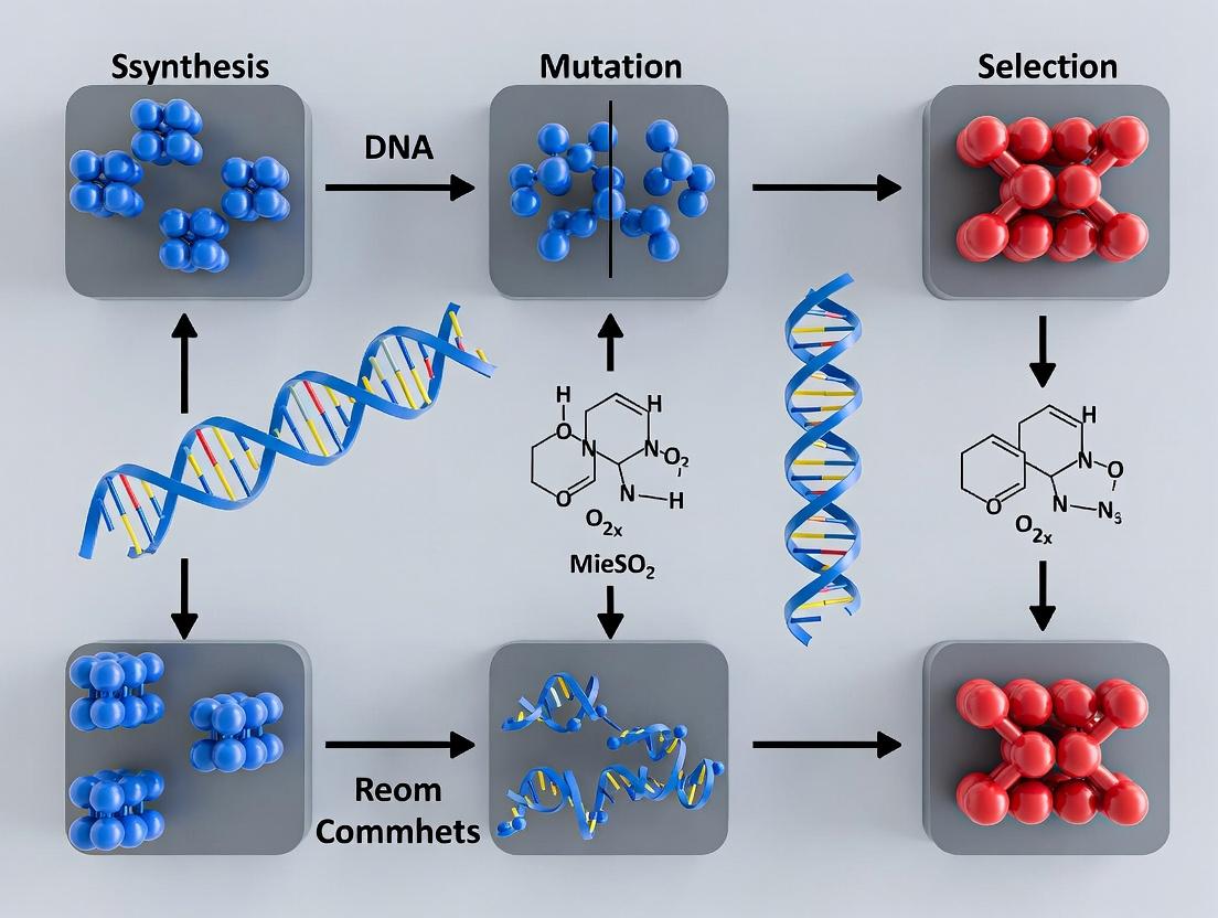
Application Notes
1. Therapeutic Enzyme Engineering for Lysosomal Storage Disorders Lysosomal enzymes like iduronate-2-sulfatase (IDS) for Hunter syndrome require engineering for improved stability at neutral pH (for bloodstream survival) and enhanced mannose-6-phosphate receptor binding for cellular uptake. DNA shuffling of human IDS with orthologs creates variant libraries screened for both catalytic activity and binding affinity.
2. Biocatalysis for Pharmaceutical Intermediate Synthesis The synthesis of chiral amines, crucial for active pharmaceutical ingredients (APIs), relies on transaminases. Wild-type enzymes often exhibit poor activity toward bulky substrates. Shuffling genes from Vibrio fluvialis and Chromobacterium violaceum generates variants capable of converting prochiral ketones to (S)-amines with >99% enantiomeric excess (ee) at industrial substrate loadings (>100 g/L).
Quantitative Data Summary: Engineered vs. Wild-Type Enzymes Table 1: Performance Metrics of Engineered Enzymes in Key Applications
| Application | Enzyme | Key Metric | Wild-Type | DNA-Shuffled Variant | Improvement Factor |
|---|---|---|---|---|---|
| Therapeutic Delivery | Iduronate-2-sulfatase (IDS) | Plasma Half-life (in vivo model) | ~3-5 minutes | ~30-40 minutes | 8-10x |
| Chiral Amine Synthesis | (S)-Transaminase | Specific Activity (on bulky substrate) | < 0.1 U/mg | 4.5 U/mg | >45x |
| Antibody Conjugation | Microbial Transglutaminase | Reaction Rate (kcat/KM) with non-native substrate | 12 M⁻¹s⁻¹ | 280 M⁻¹s⁻¹ | ~23x |
| Continuous Flow Manufacturing | Lipase B (Immobilized) | Total Turnover Number (TTN) at 60°C | 1.2 x 10⁵ | 2.1 x 10⁶ | ~17.5x |
| CAR-T Cell Therapy | Activation Caspase (safety switch) | Activation Time upon Induction | ~60 minutes | <20 minutes | 3x faster |
Experimental Protocols
Protocol 1: DNA Shuffling and Screening for Thermostable Lipases Objective: Generate a thermostable lipase variant for continuous flow biocatalysis. Materials: Parental lipase genes (LipA, LipB from thermophiles), E. coli BL21(DE3) expression system, p-nitrophenyl palmitate (pNPP) assay reagents, thermocycler.
Methodology:
- Gene Fragmentation: Digest 1 µg each of purified lipA and lipB genes with 0.15 units of DNase I in 50 µL Tris-MgCl2 buffer for 10 minutes at 25°C to yield random 50-200 bp fragments.
- Reassembly PCR: Purify fragments and assemble without primers. Use 5 µL of fragment mix in a 50 µL PCR reaction: 30 cycles of (94°C for 30s, 50°C for 30s, 72°C for 30s). This allows homologous fragments to prime each other, recombining sequences.
- Amplification: Add gene-specific primers to the reassembly product and amplify with standard PCR (30 cycles).
- Cloning & Expression: Clone shuffled library into pET-28a vector, transform into E. coli, and plate on LB/Kanamycin.
- Primary Screen (Thermal Challenge): Pick colonies into 96-deep well plates. After induction, apply a heat challenge (65°C for 30 min) to cell lysates. Assess residual activity via pNPP assay (A405 measurement).
- Secondary Screen: Re-test positive hits from primary screen under process conditions (immobilized enzyme, 60°C continuous flow). Select top variant (e.g., ShuffledLip-05 from Table 1) for characterization.
Protocol 2: Engineering Caspase-9 for Rapid-Response Safety Switches Objective: Create a faster-activating, dimerizer-dependent caspase-9 for controlled CAR-T cell ablation. Methodology:
- Library Creation: Shuffle human caspase-9 with its more active orthologs (e.g., from Danio rerio). Focus recombination on the pro-domain and linker region.
- Yeast Surface Display: Clone the shuffled library into a yeast display vector. Induce expression in Saccharomyces cerevisiae EBY100.
- FACS Screening: Label yeast with a fluorescent inhibitor probe (FITC-DEVD-fmk) to detect active caspase conformations. Use a second label for dimerizer drug (AP20187) binding. Employ FACS to isolate cells showing high FITC signal only in the presence of the dimerizer, indicating inducible activity.
- Functional Validation: Clone recovered variants into CAR-T cells. Induce with dimerizer and measure apoptosis kinetics via live-cell imaging (Annexin V staining). Select the fastest-responding variant (e.g., activation <20 min).
Visualizations
Title: DNA Shuffling and Screening Workflow for Enzyme Engineering
Title: Engineered Caspase-9 Safety Switch Activation Pathway
The Scientist's Toolkit: Key Research Reagent Solutions
Table 2: Essential Materials for DNA Shuffling & Enzyme Screening
| Item | Function & Rationale |
|---|---|
| DNase I (Grade I) | Creates random fragments of parental DNA for shuffling. Purity is critical to prevent nicking of templates. |
| PfuTurbo DNA Polymerase | High-fidelity polymerase for reassembly and amplification PCR to minimize point mutations during shuffling. |
| Yeast Surface Display Vector (pYD1) | For displaying shuffled enzyme libraries on the yeast cell surface for FACS-based screening of binding/activity. |
| Fluorescent Activity-Based Probe (ABP) | e.g., FITC-DEVD-fmk. Covalently labels active enzyme variants in live cells for functional screening. |
| p-Nitrophenyl Ester Substrates (pNP) | Chromogenic substrates (e.g., pNPP) for high-throughput kinetic assays of esterase/lipase activity in lysates. |
| Thermostable Affinity Resin (e.g., Ni-NTA) | For rapid, heat-challenge purification of His-tagged variants. Stability at 60°C+ allows screening for thermostability. |
| Dimerizer Drug (AP20187) | Chemically induces dimerization of engineered caspase-9 safety switches in cellular therapies. |
| Microfluidic Continuous-Flow Reactor | Enables true process-mimetic screening of immobilized enzyme variants under industrial conditions. |
Natural evolution, driven by random mutation and natural selection over millennia, is the foundation of biological diversity. Directed evolution, pioneered by Frances Arnold and others, harnesses these principles in the laboratory to engineer biomolecules with desired traits. This represents a fundamental conceptual leap: from observing evolution to actively designing and controlling it. In the context of enzyme engineering, this shift enables the rapid optimization of enzymes for industrial catalysis, therapeutic applications, and diagnostics. DNA shuffling, a method of in vitro homologous recombination, is a cornerstone technique that accelerates this process by mimicking sexual recombination to generate genetic diversity.
Core Principles & Quantitative Comparison
The table below contrasts the key parameters of natural and directed evolution, highlighting the efficiency gains.
Table 1: Comparative Analysis of Natural vs. Directed Evolution
| Parameter | Natural Evolution | Directed Evolution (with DNA Shuffling) |
|---|---|---|
| Time Scale | Millions of years | Weeks to months |
| Diversity Generation | Random point mutations, sexual recombination | Controlled mutagenesis (error-prone PCR, shuffling) |
| Selection Pressure | Environmental fitness (survival & reproduction) | User-defined, high-throughput screening/selection |
| Throughput | Population-level | 10^4 – 10^8 variants per cycle |
| Primary Goal | Adaptation to environment | Optimization of specific trait(s) (e.g., activity, stability) |
| Control & Direction | Undirected, stochastic | Highly directed, iterative |
Key Research Reagent Solutions
The following toolkit is essential for implementing a DNA shuffling-based directed evolution campaign.
Table 2: Essential Research Reagents for DNA Shuffling & Enzyme Engineering
| Reagent / Material | Function / Purpose |
|---|---|
| Parental Gene Templates | Heterologous genes with sequence homology for shuffling; provide the starting genetic diversity. |
| DNase I | Randomly fragments the parental genes to create a pool of small DNA fragments for reassembly. |
| dNTPs | Deoxynucleotide triphosphates; building blocks for PCR-based reassembly and amplification. |
| Taq DNA Polymerase | Thermostable polymerase for PCR reassembly and amplification. Lacks proofreading to allow minor misincorporation. |
| High-Fidelity Polymerase (e.g., Q5) | For final amplification of shuffled library with minimal errors, prior to cloning. |
| Restriction Enzymes & Ligase | For cloning the shuffled gene library into an appropriate expression vector. |
| Expression Vector & Host (E. coli) | System for expressing the library of variant enzymes. |
| Selection/Agar Plates with Antibiotic | To select for host cells containing the expression vector. |
| Substrate for Enzyme Assay | Critical for high-throughput screening; often a chromogenic, fluorogenic, or selective growth substrate. |
| Microtiter Plates (96- or 384-well) | Platform for parallel cell culture and high-throughput enzymatic assays. |
| Plate Reader | For rapid quantification of assay signals (absorbance, fluorescence) across the variant library. |
Protocols
Protocol 4.1: Basic DNA Shuffling Workflow
This protocol describes the generation of a shuffled gene library from multiple parental sequences.
Materials:
- Purified DNA of parent genes (≥ 90% homology).
- DNase I (1 U/µL), 10x DNase I buffer.
- Agarose gel electrophoresis system.
- PCR reagents: Taq polymerase, 10x buffer, dNTPs, primers.
- Gel extraction/PCR purification kit.
Procedure:
- Fragmentation: Combine 1-5 µg of total parent DNA in 100 µL of 1x DNase I buffer. Add DNase I to a final concentration of 0.15 U/µL. Incubate at 15°C for 10-20 min. Quench with 10 µL of 0.5 M EDTA and heat at 90°C for 10 min.
- Size Selection: Purify fragments and run on a 2% agarose gel. Excise fragments in the 10-50 bp size range and purify using a gel extraction kit. Elute in 30 µL water.
- Reassembly PCR: In a 100 µL PCR reaction, combine purified fragments (10-100 ng) without primers. Use a low primer extension temperature. Typical program: 95°C for 2 min; then 40-60 cycles of: 94°C for 30s, 50-55°C for 30s, 72°C for 1-2 min (no final extension). This allows fragments to prime each other based on homology, reassembling into full-length genes.
- Amplification: Dilute the reassembly product 10-fold. Perform a standard PCR using gene-specific primers that introduce restriction sites for cloning. Use a high-fidelity polymerase.
- Cloning & Library Creation: Digest the PCR product and expression vector with appropriate restriction enzymes. Ligate and transform into competent E. coli. Plate on selective media to create the library.
Protocol 4.2: High-Throughput Screening for Thermostability
This protocol outlines a common screening method for identifying shuffled enzymes with improved thermal stability.
Materials:
- Library colonies in 96-well culture blocks.
- LB media with antibiotic.
- Lysis buffer (e.g., BugBuster Master Mix).
- Assay buffer and substrate.
- Thermocycler or heat block.
- Microtiter plate reader.
Procedure:
- Expression: Inoculate library colonies into deep 96-well plates containing 1 mL of LB with antibiotic. Grow overnight at 37°C, 220 rpm.
- Induction & Harvest: Dilute cultures 1:100 into fresh media, grow to mid-log phase, induce with IPTG. Harvest cells by centrifugation after expression.
- Crude Lysate Preparation: Resuspend cell pellets in 200 µL of lysis buffer. Incubate with shaking for 20 min. Clarify by centrifugation (15 min, 4000 x g). Transfer supernatant (crude lysate) to a new plate.
- Heat Challenge: Aliquot two 50 µL samples of each lysate. Incubate one "test" plate at the challenge temperature (e.g., 55°C) for 30 min. Keep the "control" plate on ice.
- Activity Assay: Transfer 10-20 µL of heat-challenged and control lysates to a fresh 96-well assay plate. Initiate reaction by adding substrate in assay buffer. Monitor product formation kinetically using a plate reader.
- Analysis: Calculate residual activity for each variant: (Activityheated / Activityunheated) * 100%. Select clones with the highest residual activity for sequencing and re-testing.
Visualizations
DNA Shuffling Experimental Workflow
Directed Evolution Cycle with DNA Shuffling
DNA shuffling, a cornerstone of directed evolution, revolutionized enzyme engineering by mimicking sexual recombination in vitro. This method allows researchers to rapidly evolve proteins with novel or enhanced functions—such as thermostability, substrate specificity, and catalytic efficiency—which is central to industrial biocatalysis and therapeutic protein development. This article, framed within a broader thesis on DNA shuffling for enzyme engineering, details its genesis, foundational protocols, and contemporary applications, providing actionable insights for researchers.
Key Pioneers and Seminal Papers
The field was pioneered by Willem P.C. Stemmer in the early 1990s. His work established the core principle of recombining homologous gene sequences to generate diverse chimeric libraries.
Table 1: Foundational Pioneers and Seminal Publications
| Year | Key Pioneer(s) | Paper Title (Journal) | Core Contribution | Quantitative Impact (Example) |
|---|---|---|---|---|
| 1994 | Willem P.C. Stemmer | "Rapid evolution of a protein in vitro by DNA shuffling" (Nature) | Introduced DNA shuffling by random fragmentation & reassembly of a single gene. | Evolved β-lactamase: 32,000x increase in MIC for cefotaxime vs. wild-type. |
| 1998 | Willem P.C. Stemmer, et al. | "Molecular evolution by staggered extension process (StEP) in vitro recombination" (PNAS) | Introduced StEP, a simplified method using PCR without fragmentation. | Recombined Bacillus subtilis subtilisin E genes; identified variants with 6x improved organic solvent resistance. |
| 2000 | Frances H. Arnold, et al. | "Directed evolution of a thermostable esterase" (PNAS) | Applied DNA shuffling to create thermostable enzymes. | Evolved esterase: 50-60°C increase in melting temperature (Tm) after 7 rounds. |
| 2001 | Andreas Crameri, Sunghwa Kim, et al. | "Molecular breeding of viruses" (Nature Biotechnology) | Extended shuffling to whole viral genomes (e.g., adenoviruses). | Generated chimeric adenoviruses with expanded tropism; titer increased by >100-fold in target cells. |
Detailed Protocols
Protocol 1: Classical DNA Shuffling (Based on Stemmer, 1994)
Objective: Recombine a family of homologous genes to create a library of chimeric sequences.
Materials (Research Reagent Solutions):
- Target DNA: Pool of homologous genes (≥ 70% identity).
- DNase I: For random fragmentation.
- DNA Polymerase I (Klenow fragment): For fragment reassembly.
- Primers: Flanking gene-specific primers for amplification.
- dNTPs: Deoxynucleotide triphosphate mix.
- PCR Reagents: Taq DNA Polymerase, buffer, MgCl₂.
Procedure:
- Fragment Preparation: Digest 10 µg of pooled DNA with 0.15 U of DNase I in 100 µL of 50 mM Tris-HCl (pH 7.4), 10 mM MnCl₂ for 10-20 min at 25°C. Generate random fragments of 50-100 bp.
- Purification: Gel-purify fragments in the 50-100 bp range.
- Reassembly PCR: In a 100 µL reaction without added primers, combine fragments (10-100 ng/µL), 0.2 mM dNTPs, 2.5 U of DNA Polymerase I Klenow fragment, in standard buffer. Thermocycle: 94°C for 2 min; then 35-45 cycles of: 94°C for 30 sec, 50-55°C for 30 sec, 72°C for 30 sec + 5 sec/cycle.
- Amplification: Use 1-5 µL of reassembly product as template in a standard 50 µL PCR with flanking primers to amplify full-length chimeric genes.
- Cloning & Screening: Clone into an appropriate expression vector and screen/select for desired phenotypes.
Protocol 2: Staggered Extension Process (StEP) Recombination (Based on Stemmer, 1998)
Objective: Simplify in vitro recombination using very short annealing/extension cycles.
Procedure:
- Template Preparation: Mix 10-100 ng of each homologous DNA template.
- StEP Cycling: In a 50 µL PCR, use gene-flanking primers. Thermocycle: 94°C for 3 min; then 80-100 cycles of: 94°C for 30 sec, 50-55°C for 5-15 sec. Critical: The extension time is too short to complete full-length synthesis, prompting template switching.
- Final Extension: After cycling, perform a final 5 min extension at 72°C.
- Amplify & Clone: Use product as template for a final PCR with flanking primers, then clone and screen.
Visualizations
Title: Classic DNA Shuffling Workflow
Title: StEP Recombination Logic
The Scientist's Toolkit: Essential Reagents & Materials
Table 2: Key Research Reagent Solutions for DNA Shuffling
| Reagent/Material | Function in DNA Shuffling | Key Consideration |
|---|---|---|
| DNase I (RNA-free) | Creates random DNA fragments for classic shuffling. | Use Mn²⁺ buffer for random cleavage; concentration and time are critical for optimal fragment size. |
| DNA Polymerase I (Klenow) | Reassembles fragments in primerless PCR. | Lacks 5'→3' exonuclease, preferred for seamless reassembly. |
| High-Fidelity DNA Polymerase (e.g., Q5, Phusion) | For final amplification of shuffled library. | Minimizes introduction of point mutations during PCR. |
| dNTP Mix (25 mM each) | Building blocks for DNA synthesis during reassembly and PCR. | Use high-quality, pH-balanced stocks to prevent degradation. |
| Homologous Gene Pool (≥70% identity) | Substrate for recombination. | Higher identity yields more crossovers and functional chimeras. |
| Gel Extraction Kit | Purification of DNA fragments (50-100 bp) or final products. | Essential for removing primers, enzymes, and incorrect fragments. |
| Expression Vector & Competent Cells | Cloning and expressing the shuffled gene library. | Vector must be compatible with host (e.g., E. coli) for high-throughput screening. |
Article Context
This Application Note details the protocol for Random Fragmentation and Reassembly PCR (RFR-PCR), a foundational DNA shuffling method central to enzyme engineering research. This technique facilitates the in vitro directed evolution of proteins by generating extensive genetic diversity through recombination of homologous parent genes.
Random Fragmentation and Reassembly PCR is a two-step process that mimics sexual recombination. First, a pool of related parent genes is randomly fragmented. Second, these fragments are reassembled into full-length chimeric genes through a primerless PCR, where fragments prime each other based on homology.
Table 1: Key Quantitative Parameters for Optimized RFR-PCR
| Parameter | Typical Range | Optimal Value | Function & Impact |
|---|---|---|---|
| DNAse I Concentration | 0.15 - 0.30 U/µg DNA | 0.20 U/µg DNA | Controls fragment size distribution. Lower yields larger fragments, higher yields smaller fragments. |
| Fragment Size Range | 50 - 200 bp | 50 - 100 bp | Smaller fragments increase crossover frequency and diversity. |
| Primerless PCR Cycles | 30 - 50 cycles | 40 cycles | Drives reassembly of fragments into full-length genes. |
| Reassembly PCR Template | 100 - 500 ng fragments | 200 ng fragments | Amount of fragmented DNA used in the primerless reassembly step. |
| Final Amplification PCR Cycles | 20 - 30 cycles | 25 cycles | Amplifies reassembled full-length products for cloning. |
| Homology Requirement | ≥ 15 bp | 20 - 50 bp | Minimum region of sequence identity for fragments to anneal and prime synthesis. |
Table 2: Comparison of DNA Shuffling Method Attributes
| Method | Crossover Control | Family Size Limit | Required Homology | Best For |
|---|---|---|---|---|
| RFR-PCR (Stemmer 1994) | Random, low | High (many parents) | Moderate to High (>70%) | Recombining highly homologous sequences (>80% identity). |
| ITCHY | Controlled, sequential | Two | None | Creating fusions of unrelated genes or domains. |
| SHIPREC | Controlled, sequential | Two | None | Generating single-crossover libraries from low-homology parents. |
| Staggered Extension (StEP) | Random, small tracts | Moderate | Moderate | Quick, single-pot recombination of 2-4 parents. |
Detailed Experimental Protocols
Protocol 2.1: Random Fragmentation of Parental DNA using DNAse I
Objective: To generate a pool of small, random DNA fragments from parental gene sequences.
Materials: See "The Scientist's Toolkit" below. Procedure:
- Prepare DNA Pool: Mix 2-5 µg of purified parental DNA (PCR products or plasmid) in a total volume of 50 µL of 1x DNAse I buffer (e.g., 50 mM Tris-Cl pH 7.6, 10 mM MnCl₂). Mn²⁺ is critical for generating random double-strand breaks.
- Titrate DNAse I (Critical): Perform a pilot digestion. Add varying amounts of DNAse I (e.g., 0.15, 0.20, 0.25 U/µg DNA) to separate tubes. Incubate at 25°C for 10 minutes.
- Stop Reaction: Terminate digestion by adding 5 µL of 0.5 M EDTA (pH 8.0) and heating at 80°C for 10 minutes.
- Analyze Fragment Size: Run 10 µL of each digestion on a 2-3% agarose gel. The optimal digest should yield a smear centered around 50-100 bp.
- Purify Fragments: Pool the optimal digest(s) and purify using a silica-membrane-based PCR cleanup kit. Elute in 30 µL of nuclease-free water. Quantify by spectrophotometry.
Protocol 2.2: Primerless Reassembly PCR
Objective: To reassemble random fragments into full-length chimeric genes via homology-driven primer extension.
Procedure:
- Set Up Reassembly Reaction:
- Template: 100-200 ng of purified fragments.
- dNTPs: 0.2 mM each.
- Taq DNA Polymerase: 2.5 U per 50 µL reaction.
- Buffer: Standard 1x PCR buffer with MgCl₂.
- No primers are added.
- Total Volume: 50 µL.
- Run Reassembly PCR Program:
- Step 1: Denaturation: 95°C for 2 min.
- Step 2: Reassembly Cycles (40 cycles):
- 95°C for 30 sec (denaturation).
- 50-60°C for 30 sec (annealing). Start at 5-10°C below the Tm of the parent genes. Optimize temperature to favor correct homology alignment.
- 72°C for 30 sec (extension). Time is short to promote fragment priming rather than long synthesis.
- Step 3: Final Extension: 72°C for 5 min.
- Hold at 4°C.
- Analyze Product: Run 5 µL on a 1% agarose gel. A successful reassembly shows a faint smear culminating in a band at the expected full-length gene size.
Protocol 2.3: Amplification of Reassembled Products
Objective: To amplify the pool of full-length, reassembled genes for subsequent cloning.
Procedure:
- Dilute Reassembly Product: Use 1 µL of the reassembly PCR product as template in a 50 µL standard PCR.
- Set Up Amplification PCR:
- Template: 1 µL reassembly product.
- Gene-Specific Primers: 0.5 µM each (forward and reverse, containing desired restriction sites for cloning).
- dNTPs: 0.2 mM each.
- High-Fidelity DNA Polymerase (e.g., Phusion): 1 U per 50 µL reaction.
- Buffer: As specified for the polymerase.
- Run Amplification PCR Program:
- Initial Denaturation: 98°C for 30 sec.
- Amplification (25 cycles): 98°C for 10 sec, Primer Tm for 20 sec, 72°C for 30 sec/kb.
- Final Extension: 72°C for 5 min.
- Purify and Clone: Gel-purify the amplified band at the correct size. Digest with appropriate restriction enzymes and clone into your expression vector for transformation and library screening.
Visualization of Workflow and Mechanism
Title: RFR-PCR Workflow from Parent Genes to Library
Title: Reassembly PCR Mechanism: Homology-Driven Recombination
The Scientist's Toolkit
Table 3: Essential Research Reagents for RFR-PCR
| Reagent / Material | Function & Critical Notes | Example Product/Catalog |
|---|---|---|
| Parent DNA Sequences | Highly homologous genes (>70% identity) serving as diversity sources. Must be purified (PCR or plasmid). | N/A |
| Deoxyribonuclease I (DNase I) | Enzyme for random double-strand fragmentation. Must be used with Mn²⁺ buffer for random cuts. | RNase-free DNase I (e.g., Thermo Scientific #EN0521) |
| PCR Purification Kit | For cleaning up fragmented DNA and final PCR products. Silica-membrane based. | QIAquick PCR Purification Kit (Qiagen) |
| Thermostable DNA Polymerase | 1) Standard Taq for primerless reassembly. 2) High-fidelity polymerase for final amplification to avoid extra mutations. | Taq DNA Pol (NEB); Phusion HF (Thermo) |
| Gene-Specific Primers | Designed with restriction sites for cloning the final shuffled library. | Custom oligonucleotides |
| Cloning Vector & Competent Cells | For library construction and expression. High-efficiency cells are crucial for library size. | pET vector series; NEB 5-alpha or similar |
| Agarose Gel Electrophoresis System | To analyze fragment size after DNase I digestion and to purify the final reassembled product. | Standard horizontal gel system |
Application Notes: DNA Shuffling in Enzyme Engineering
DNA shuffling is an in vitro directed evolution method that mimics natural recombination to accelerate the evolution of proteins. It addresses the limitations of point mutagenesis by enabling the recombination of beneficial mutations from multiple parent genes, facilitating the exploration of vast sequence spaces. This protocol series details the core methodology and its application for evolving enzymes with improved properties such as thermostability, catalytic activity, and substrate specificity for pharmaceutical and industrial biocatalysis.
Table 1: Quantitative Outcomes from Recent DNA Shuffling Studies (2022-2024)
| Enzyme Target | Parent Genes/ Variants | Shuffling Method | Key Improved Trait | Fold Improvement | Reference (Type) |
|---|---|---|---|---|---|
| PET Hydrolase | 4 thermostable variants | Family shuffling | Melting Temp (Tm) | +12.5°C | Nat. Commun. (2023) |
| CYP450 Monooxygenase | 3 homologs from fungi | SCRATCHY | Total Turnover Number | 8.7x | ACS Catal. (2022) |
| β-Lactamase | Error-prone PCR library | StEP (Staggered Extension) | MIC of Ampicillin | 32,000x | Proc. Natl. Acad. Sci. (2023) |
| Transaminase | 5 bacterial genes | ITCHY (Incremental Truncation) + Shuffling | Stereoselectivity (ee) | >99% (from 78%) | Angew. Chem. (2024) |
| AAV Capsid | 7 serotype libraries | DOGS (DNA-family shuffling) | Liver Tropism (vs. AAV9) | 45x higher | Cell (2023) |
Protocol 1: Standard Family DNA Shuffling for Chimeric Enzyme Libraries
Objective: To generate a diverse library of chimeric enzymes by recombining homologous genes from different species or designed variants.
Materials & Reagents:
- DNA Template: Pool of parent genes (≥70% sequence identity, 50-100 ng each).
- Enzymes: DNase I (for random fragmentation), DNA Polymerase I (Klenow fragment, for reassembly), Thermostable DNA Polymerase (e.g., Taq or Q5, for amplification).
- Buffers: DNase I digestion buffer, PCR assembly buffer.
- Purification Kits: Gel extraction kit, PCR purification kit.
- Cloning Vector: Restriction enzyme-linearized plasmid backbone.
- Assembly Mix: Gibson Assembly Master Mix or similar.
Procedure:
- Fragment Generation: Combine parent genes. Add DNase I (0.15 U/µg DNA) in 1x digestion buffer with 2.5 mM MnCl₂. Incubate at 15°C for 10-20 min to generate random fragments (10-50 bp). Heat-inactivate at 80°C for 10 min.
- Reassembly PCR: Without purification, add dNTPs (0.2 mM), primers (0.1 µM flanking gene ends), and Taq polymerase. Run reassembly PCR: 94°C for 2 min; then 35 cycles of [94°C for 30s, 50-55°C for 30s, 72°C for 1 min/kb] with no primers in the first 5-10 cycles to allow fragment priming; then add primers for final amplification.
- Library Amplification & Cloning: Purify reassembly product. Reamplify full-length genes with high-fidelity polymerase using flanking primers containing overlaps for cloning. Purify PCR product. Use Gibson Assembly to clone into linearized expression vector.
- Transformation & Screening: Transform assembly into competent E. coli (e.g., NEB 10-beta). Plate on selective media. Pick colonies for high-throughput screening based on desired enzyme activity.
The Scientist's Toolkit: Key Reagents for DNA Shuffling
| Reagent / Material | Function & Critical Role in Protocol |
|---|---|
| DNase I (Grade I) | Creates random double-stranded breaks in parent DNA to generate a pool of short fragments for recombination. Mn²⁺ condition is crucial for random cleavage. |
| Klenow Fragment (exo-) | Used in the reassembly step to fill gaps and create nicked double-stranded DNA from overlapping fragments, prior to full PCR amplification. |
| Gibson Assembly Master Mix | Enables seamless, one-pot cloning of the shuffled gene library into an expression vector, essential for high-efficiency library construction. |
| NGS Library Prep Kit | For deep sequencing of input and output populations to analyze crossover frequency, identify consensus sequences, and map beneficial mutations. |
| Phusion or Q5 High-Fidelity Polymerase | Used for final amplification of shuffled full-length genes to minimize the introduction of spurious point mutations during PCR. |
| Automated Colony Picker | Enables rapid transfer of thousands of bacterial colonies to microtiter plates for high-throughput enzymatic screening assays. |
Protocol 2: Staggered Extension Process (StEP) for Recombination
Objective: A simplified shuffling method performed in a single thermocycler reaction, suitable for recombining closely related sequences.
Materials & Reagents: Parent DNA templates, high-fidelity DNA polymerase, dNTPs, forward and reverse primers.
Procedure:
- Template Mix: Combine equimolar amounts (10-50 ng each) of parent DNA templates in a standard PCR mix.
- StEP Cycling Program: Run 80-100 cycles of a short denaturation (94°C for 20s) followed by an extremely short annealing/extension (45-55°C for 5-10s). This abbreviated extension time causes the polymerase to repeatedly extend and dissociate, switching templates and creating chimeric sequences.
- Full-Length Product Isolation: After cycling, run the product on an agarose gel. Excise and purify the band corresponding to the full-length gene.
- Final Amplification & Cloning: Reamplify the purified full-length product with a standard PCR protocol. Clone and screen as in Protocol 1.
Diagram Title: DNA Shuffling by Fragment Reassembly Workflow
Diagram Title: StEP Recombination Mechanism
DNA shuffling is a directed evolution technique used to engineer enzymes with improved properties (e.g., stability, activity, substrate specificity). The success of this method is fundamentally dependent on two critical initial steps: generating high-quality genetic diversity and selecting optimal parent sequences. These prerequisites set the stage for effective recombination and subsequent screening. This protocol outlines the contemporary methodologies for these foundational stages within a research thesis focused on advancing enzyme engineering for therapeutic and industrial applications.
Table 1: Quantitative Metrics for Parent Gene Selection in DNA Shuffling
| Metric | Target Range / Value | Measurement Method | Rationale |
|---|---|---|---|
| Sequence Identity | 60-90% | Multiple Sequence Alignment (e.g., Clustal Omega) | Ensures sufficient homology for effective recombination while maintaining diversity. |
| Functional Diversity | ≥ 40% variance in key parameter (e.g., kcat/Km) | Enzymatic assays under standardized conditions | Guarantees that shuffling pools beneficial mutations from distinct functional backgrounds. |
| Thermostability (Tm) | Spread of ≥ 10°C | Differential scanning fluorimetry (DSF) | Allows recombination to blend stability traits from mesophilic and thermophilic parents. |
| Expression Level | > 10 mg/L in host system | SDS-PAGE / spectrophotometry | Ensures parent genes are expressible, avoiding library bias from poor expression. |
| Number of Parents | 4-8 genes | N/A | Optimizes diversity and screening load; too few limits diversity, too many dilutes beneficial mutations. |
Table 2: Common Gene Diversity Generation Methods (2024 Benchmarks)
| Method | Avg. Mutation Rate (%) | Library Size (Variants) | Key Application |
|---|---|---|---|
| Error-Prone PCR (epPCR) | 0.1 - 2.0 | 10⁴ - 10⁶ | Introducing point mutations across entire gene. |
| Site-Saturation Mutagenesis | 100 at chosen codons | 10² - 10³ per site | Exploring all amino acid possibilities at specific residues. |
| Oligonucleotide-Directed Mutagenesis | User-defined | 10³ - 10⁵ | Introducing targeted blocks of diversity in specific regions. |
| Gene Synthesis (Fragment-Based) | Fully designed | 10³ - 10⁵ | Creating synthetic gene libraries with pre-calculated diversity. |
Experimental Protocols
Protocol 1: In Silico Parent Selection and Analysis
Objective: To computationally select and characterize parent genes for DNA shuffling.
Materials:
- Homologous gene sequences (from NCBI, metagenomic databases, or in-house libraries).
- Bioinformatics software: Clustal Omega, BLAST, Pymol, or similar.
- Research Reagent Solutions: See Toolkit (Table 3).
Method:
- Sequence Acquisition & Alignment:
- Gather candidate gene sequences (DNA and protein) encoding the target enzyme from diverse organisms.
- Perform multiple sequence alignment using Clustal Omega. Calculate pairwise sequence identity.
- Phylogenetic Analysis:
- Construct a neighbor-joining tree to visualize evolutionary relationships. Select parents from distinct clades to maximize functional diversity.
- Structural Mapping:
- If available, map variable regions onto a 3D protein structure (e.g., using Pymol). Prioritize parents with variations in active site loops, substrate channels, or subunit interfaces.
- Computational Fitness Prediction:
- Use tools like FoldX or Rosetta to estimate stability changes (ΔΔG) of parent variants. Filter out parents with predicted severely destabilizing mutations.
- Final Selection:
- Apply criteria from Table 1. Select 4-8 parent genes that collectively maximize coverage of sequence space and functional attributes.
Protocol 2: Generation of Diversity via Tunable Error-Prone PCR (epPCR)
Objective: To create a mutagenized library of a single parent gene to supplement shuffling diversity.
Materials:
- High-fidelity DNA polymerase (e.g., Q5).
- Mutagenic polymerase blend (e.g., Mutazyme II).
- dNTPs, including biased dNTP pools (e.g., increased dATP/dTTP).
- Target plasmid DNA (50 ng/µL).
- Thermostable PCR machine.
- PCR purification kit.
Method:
- Reaction Setup (50 µL):
- Template DNA: 10-50 ng.
- Forward/Reverse primers (flanking gene): 0.5 µM each.
- Mutazyme II buffer: 1X.
- Biased dNTP mix: 0.2 mM dGTP, 0.2 mM dCTP, 1.0 mM dATP, 1.0 mM dTTP.
- Mutazyme II polymerase: 1 unit.
- Adjust with nuclease-free water.
- Thermocycling:
- Initial denaturation: 95°C for 2 min.
- 30 cycles: 95°C for 30 sec, 55°C (Tm-specific) for 30 sec, 72°C for 1 min/kb.
- Final extension: 72°C for 5 min.
- Product Analysis & Purification:
- Run 5 µL on agarose gel to confirm amplicon size.
- Purify the remaining PCR product using a spin column kit.
- Quantify DNA concentration via spectrophotometry.
Visualization
Diagram 1: Parent Selection Workflow for DNA Shuffling
Diagram 2: Gene Diversity Generation Pathways
The Scientist's Toolkit: Research Reagent Solutions
Table 3: Essential Materials for Gene Diversity & Parent Selection
| Item | Function & Application | Example Product/Brand |
|---|---|---|
| High-Fidelity Polymerase | Amplifying parent genes without introducing errors for cloning. | Q5 (NEB), Phusion (Thermo) |
| Mutagenic Polymerase Blend | Performing error-prone PCR with adjustable mutation rates. | Mutazyme II (Agilent), Taq Pol (low fidelity) |
| Biased dNTP Mix | Skewing nucleotide incorporation to increase mutation frequency in epPCR. | Custom mix from Jena Bioscience |
| Next-Gen Sequencing Kit | Validating library diversity and mutation rates pre-shuffling. | Illumina MiSeq, Oxford Nanopore |
| Thermal Shift Dye | Measuring protein thermostability (Tm) of parent variants via DSF. | SYPRO Orange (Thermo) |
| Cloning/Assembly Master Mix | Efficiently assembling shuffled gene fragments into expression vectors. | Gibson Assembly Master Mix (NEB), Golden Gate Assembly kits |
| Expression Host Cells | Testing parent gene expressibility and function (prokaryotic/eukaryotic). | E. coli BL21(DE3), P. pastoris strains |
| Activity Assay Substrate | Quantifying functional diversity of parent enzymes via kinetic assays. | Fluorogenic/Chromogenic substrates (e.g., pNPP for phosphatases) |
Protocols and Applications: A Step-by-Step Guide to Shuffling Enzymes
This application note details a core methodology within the broader thesis on DNA shuffling for directed enzyme evolution. DNA shuffling, or sexual PCR, is a powerful technique for in vitro recombination of homologous genes, generating diverse chimeric libraries for screening improved protein variants. This protocol outlines the standard workflow from random fragmentation of parental genes to their reassembly into full-length sequences.
Experimental Protocols
Protocol 1: DNase I Random Fragmentation
Objective: To generate a pool of small random DNA fragments (10-50 bp) from parental gene templates for subsequent shuffling.
Materials:
- Purified parental DNA (e.g., homologous genes or gene family).
- DNase I (RNase-free, 1 U/µL).
- 10x DNase I Reaction Buffer (typically 100 mM Tris-HCl, pH 7.5, 25 mM MgCl₂, 5 mM CaCl₂).
- Stop Solution (20 mM EGTA, pH 8.0).
- Thermo-cycler or water bath.
Method:
- In a 0.2 mL PCR tube, combine 1-2 µg of pooled parental DNA, 5 µL of 10x DNase I Buffer, and nuclease-free water to a final volume of 48 µL.
- Pre-warm the mixture to the desired digestion temperature (typically 15-25°C) for 2 minutes.
- Add 2 µL of a freshly diluted DNase I solution (diluted in cold nuclease-free water to a concentration of 0.015 U/µL).
- Incubate at the pre-warmed temperature for exactly 10 minutes. Critical: Time and enzyme concentration must be optimized to yield the desired fragment size range.
- Immediately stop the reaction by adding 5 µL of Stop Solution (20 mM EGTA) and heating at 80°C for 10 minutes.
- Purify the fragments using a standard DNA clean-up kit or gel extraction.
Protocol 2: Reassembly PCR (Assembly)
Objective: To reassemble the small random fragments into full-length chimeric genes via a primerless PCR.
Materials:
- Purified DNA fragments (from Protocol 1).
- High-fidelity DNA Polymerase (e.g., Pfu Turbo, Phusion) with corresponding 10x Buffer.
- 10 mM dNTP mix.
- Nuclease-free water.
Method:
- In a 0.2 mL PCR tube, set up the following reaction:
- DNA fragments (10-100 ng): variable.
- 10x Polymerase Buffer: 5 µL.
- dNTP mix (10 mM each): 1 µL.
- High-fidelity DNA Polymerase (2 U/µL): 1 µL.
- Nuclease-free water to 50 µL.
- Run the following thermocycling program:
- Step 1: 95°C for 2 min (initial denaturation).
- Step 2: 94°C for 30 sec.
- Step 3: 50-55°C for 30 sec. Note: Annealing temperature may require optimization based on fragment homology.
- Step 4: 72°C for 1 min per kb of expected full-length product.
- Step 5: Repeat Steps 2-4 for 35-45 cycles. Fragments prime on each other and extend.
- Step 6: 72°C for 7 min (final extension).
- Analyze 5 µL of the product by agarose gel electrophoresis. A smear culminating in a band of the expected full-length size should be visible.
- Purify the full-length product using a PCR clean-up kit.
Protocol 3: Amplification of Shuffled Library
Objective: To amplify the reassembled full-length products using external primers for downstream cloning.
Materials:
- Purified reassembly product (from Protocol 2).
- Forward and Reverse gene-specific primers (with appropriate restriction sites for cloning).
- High-fidelity DNA Polymerase and dNTPs.
Method:
- Set up a standard PCR using 1-5 µL of the purified reassembly product as template.
- Use the gene-specific primers and an annealing temperature optimized for them.
- Run for 20-25 cycles to avoid introducing additional mutations.
- Gel-purify the amplified product of the correct size.
- Digest with appropriate restriction enzymes and clone into the desired expression vector for library construction and screening.
Data Presentation
Table 1: Optimization of DNase I Digestion for Fragment Size Control
| DNase I Concentration (U/µL) | Incubation Time (min) | Temperature (°C) | Average Fragment Size (bp) | Ideal for Shuffling? (Y/N) |
|---|---|---|---|---|
| 0.03 | 10 | 25 | 20-30 | N (Too small) |
| 0.015 | 10 | 25 | 30-50 | Y |
| 0.015 | 5 | 25 | 50-100 | N (Too large) |
| 0.015 | 10 | 15 | 40-60 | Y |
| 0.0075 | 10 | 25 | 80-150 | N (Too large) |
Table 2: Critical Parameters for Reassembly PCR Success
| Parameter | Recommended Setting | Impact of Deviation |
|---|---|---|
| Fragment Concentration | 10-100 ng/50 µL rxn | Low: No product. High: Mismatched annealing. |
| Cycle Number | 35-45 | Low: Incomplete assembly. High: Excessive mutations. |
| Annealing Temperature | 50-55°C | High: Low yield. Low: Non-homologous recombination. |
| Polymerase Type | High-fidelity (e.g., Pfu) | Taq polymerase introduces excess point mutations. |
| Extension Time | 1 min/kb | Insufficient time leads to truncated products. |
Visualizations
DNA Shuffling Core Workflow
Fragment Recombination Mechanism
The Scientist's Toolkit
Table 3: Essential Research Reagent Solutions for DNA Shuffling
| Reagent / Material | Function in Workflow | Critical Notes |
|---|---|---|
| DNase I (RNase-free) | Creates random double-stranded breaks in parental DNA to generate fragment library. | Must be titrated carefully; use low concentration (e.g., 0.015 U/µL) for small fragments. |
| EGTA Stop Solution | Chelates Mg²⁺ and Ca²⁺ ions, irreversibly inactivating DNase I. | Essential for obtaining reproducible fragment sizes. |
| High-Fidelity DNA Polymerase (e.g., Pfu, Phusion) | Catalyzes the primerless reassembly PCR and final amplification with high accuracy. | Reduces introduction of spurious point mutations during recombination. |
| DNA Clean-up & Gel Extraction Kits | Purifies fragments after digestion and reassembled products before amplification. | Removes enzymes, salts, and size-selects DNA to improve downstream efficiency. |
| Gene-Specific Primers with Restriction Sites | Flank the shuffled gene for amplification and facilitate directional cloning into expression vectors. | Should be designed to anneal outside the variable region being shuffled. |
| Thermostable Pyrophosphatase (optional) | Degrades inorganic pyrophosphate (PPi) produced during PCR. | Can improve yield of long products in reassembly PCR by preventing inhibition. |
Application Notes
Within the broader thesis on DNA shuffling for enzyme engineering, classical homology-dependent shuffling methods face limitations with sequences of low identity (<70%), risking low crossover frequency and limited diversity. The modern variations discussed herein—Staggered Extension Process (StEP), Incremental Truncation for the Creation of Hybrid Enzymes (ITCHY), and Sequence Homology-Independent Protein Recombination (SHIPREC)—circumvent this by reducing or eliminating the reliance on sequence homology. They are particularly suited for:
- Engineering enzymes from distinct evolutionary families.
- Recombining genes with very low sequence identity.
- Creating focused, functional hybrid libraries when structure-function relationships are partially known.
| Method | Core Principle | Homology Requirement | Typical Library Size | Key Advantage | Primary Challenge |
|---|---|---|---|---|---|
| StEP | PCR with extremely short annealing/extension times, causing template switching. | Moderate to High (≥60%) | 10^5 – 10^6 | Simple PCR-based protocol; generates multi-crossovers. | Limited control over crossover points; requires some homology. |
| ITCHY | Controlled exonuclease digestion to create truncated fragments, ligated to form single-crossover hybrids. | None | 10^3 – 10^4 | Truly sequence-independent; creates precise 1:1 fusions. | Libraries contain non-full-length clones; limited to single crossovers. |
| SHIPREC | Random fragmentation of genes, size selection, and ligation to create fusion libraries. | None | 10^4 – 10^5 | Creates N- to C-terminal fusions from diverse parents; preserves reading frame. | Requires careful fragment size selection; can be technically demanding. |
Experimental Protocols
Protocol 1: Staggered Extension Process (StEP) for Chimeric Enzyme Library Creation
Objective: To recombine two or more parental enzyme genes (>60% identity) via template switching during PCR.
Materials:
- Template DNA: Plasmid DNA (10-50 ng each) containing parental genes.
- Primers: Forward and reverse primers flanking the gene coding sequence.
- PCR Mix: Thermostable DNA polymerase (e.g., Taq), dNTPs, MgCl₂, corresponding PCR buffer.
- Thermocycler.
Procedure:
- Set up a standard 50 µL PCR reaction containing templates, primers, dNTPs (200 µM each), MgCl₂ (1.5 mM), and polymerase (1.25 U).
- Perform the StEP cycling program:
- Initial Denaturation: 94°C for 2 min.
- Cycles (100x):
- Denaturation: 94°C for 30 sec.
- Annealing/Extension: 55°C for 5-15 sec. (Critical: This short time prevents full extension, promoting template switching.)
- Perform a final extension at 72°C for 5 min.
- Purify the PCR product using a gel extraction or PCR cleanup kit.
- Clone the shuffled library into an appropriate expression vector using restriction digestion/ligation or a seamless cloning method (e.g., Gibson Assembly).
- Transform into competent E. coli cells and plate for library screening/selection.
Protocol 2: ITCHY for Sequence-Independent Single-Crossover Hybrids
Objective: To create a comprehensive library of single-crossover fusions between two unrelated enzyme genes.
Materials:
- Linearized DNA: Parental genes A and B in separate vectors, linearized at the desired fusion junction (e.g., via restriction digest).
- Exonuclease III: For controlled 3'→5' digestion.
- Aliquot Tubes: Pre-filled with stop solution (e.g., phenol-chloroform) for timed digestion points.
- Klenow Fragment & dNTPs: For blunting ends.
- T4 DNA Ligase: For ligating complementary truncations.
- Agarose Gel & Size-Selection Equipment.
Procedure:
- Truncation: Set up two separate Exonuclease III digestions for Gene A and Gene B. Remove aliquots at fixed time intervals (e.g., every 30 seconds) into stop solution to generate a pool of fragments truncated to varying lengths.
- Blunting: Treat pooled fragments from each gene with Klenow fragment and dNTPs to create blunt ends.
- Ligation: Mix equimolar amounts of blunted Gene A and Gene B fragments. Ligate with T4 DNA Ligase. This creates a library of A-B fusions where the junction point varies.
- Size Selection: Run the ligation product on an agarose gel. Excise and purify DNA corresponding to the size of a full-length hybrid gene (length of Gene A + Gene B).
- Cloning and Expression: Amplify the size-selected library by PCR, clone into an expression vector, and transform into E. coli for screening.
Visualizations
StEP Recombination PCR Workflow
ITCHY Library Construction Protocol
The Scientist's Toolkit: Key Research Reagent Solutions
| Reagent/Material | Function in Modern Shuffling | Example/Notes |
|---|---|---|
| Thermostable DNA Polymerase | Catalyzes primer extension in StEP PCR; fidelity can influence mutation rate. | Taq polymerase (low fidelity, promotes diversity), Phusion (high fidelity). |
| Exonuclease III | Catalyzes the controlled 3'→5' truncation of dsDNA for ITCHY library construction. | Critical for generating incremental truncation pools. Requires linear dsDNA with 3'-recessed or blunt ends. |
| T4 DNA Ligase | Joins compatible ends of DNA fragments during ITCHY and SHIPREC library assembly. | Essential for creating covalent bonds between truncated or fragmented gene segments. |
| Klenow Fragment (exo-) | Fills in 5'-overhangs and removes 3'-overhangs to create blunt-ended DNA for ITCHY ligation. | Used after exonuclease digestion to prepare fragments for ligation. |
| Size-Selective Gel Extraction Kit | Purifies DNA fragments of a specific size range (e.g., full-length hybrids) from agarose gels. | Crucial for ITCHY/SHIPREC to remove non-productive ligation products. |
| Seamless Cloning Master Mix | Efficiently clones shuffled PCR products into expression vectors without restriction sites. | Gibson Assembly, In-Fusion cloning. Speeds up library construction from StEP products. |
| High-Efficiency Competent Cells | Ensures maximum transformation efficiency for comprehensive library representation. | E. coli strains like NEB 10-beta or MegaX DH10B T1R (≥10^9 CFU/µg). |
Application Notes: Context within DNA Shuffling for Enzyme Engineering
The efficacy of DNA shuffling as a directed evolution method hinges on the strategic construction of the mutant library. This process recombines homologous gene fragments from multiple parent sequences, introducing diversity through both crossover events and point mutations during PCR amplification. The core challenge is to generate a library that maximizes functional diversity while remaining within the screening or selection capacity of the available assay. A poorly designed library can be overwhelmingly large with low functional yield, rendering the process inefficient and costly.
The key parameters for strategic design are:
- Size: The total number of unique variants in the library.
- Diversity: The sequence space coverage, including the number of crossovers per gene and mutation rate.
- Screenability: The fraction of the library that can be realistically assayed for the desired function.
Table 1: Quantitative Parameters for Strategic Library Design
| Parameter | Typical Target Range | Impact on Library | Experimental Control |
|---|---|---|---|
| Library Size | 10^4 – 10^8 variants | Defines screening burden. | Adjusted via transformation efficiency & DNA input. |
| Sequence Diversity (Homology) | 70-95% between parents | Higher homology increases recombination frequency. | Parent gene selection. |
| Mutation Rate | 0.05-0.7% per nucleotide | Introduces beneficial point mutations; too high yields non-functional variants. | Controlled by PCR conditions (e.g., Mn2+ concentration, error-prone PCR cycles). |
| Crossovers per Gene | 1-4 per kb per shuffle | Increases combinatorial diversity. | Influenced by fragment size and homology. |
| Functional Fraction | Often <0.1% of library | Determines number of clones to screen for a hit. | Optimized by using high-quality, functionally validated parents. |
Experimental Protocols
Protocol 1: Standard DNA Shuffling and Library Construction
Objective: To create a shuffled library from multiple parent genes encoding homologous enzymes.
Materials: Parent plasmid DNA, DNase I, DNA purification kit, Taq DNA Polymerase (no proofreading), dNTPs, primers flanking gene sequence, appropriate E. coli expression strain, recovery media, selective agar plates.
Procedure:
- Gene Fragmentation: Combine 1-5 µg of pooled parent DNA in 100 µL of DNase I digestion buffer (e.g., 50 mM Tris-HCl, pH 7.4, 10 mM MnCl2). Add 0.15 U of DNase I per µg DNA and incubate at 15-25°C for 5-20 minutes. Target fragment sizes of 50-200 bp.
- Purification: Gel-purify fragments in the target size range.
- Reassembly PCR: Perform a PCR without primers. Use 0.2-2 µg of purified fragments in a 100 µL reaction with standard Taq buffer, 0.2 mM dNTPs, and 2.5 U of Taq polymerase. Cycle: 94°C for 2 min; then 35-45 cycles of [94°C for 30 sec, 50-60°C for 30 sec, 72°C for 30-60 sec]; final 72°C for 5 min. Fragments prime each other based on homology and reassemble into full-length genes.
- Amplification: Use 1-5 µL of the reassembly product as template in a standard PCR with gene-flanking primers to amplify the full-length shuffled genes.
- Cloning & Transformation: Digest the PCR product and vector with appropriate restriction enzymes. Ligate and transform into a competent expression host (e.g., E. coli BL21(DE3)). Plate on selective media to yield the primary library.
- Library Titering: Count colonies from a serial dilution of the transformation to calculate total library size (cfu/mL × total volume).
Protocol 2: Staggered Extension Process (StEP) for Recombination
Objective: A simplified shuffling method that combines fragmentation and reassembly in a single PCR reaction.
Materials: Parent plasmid DNA, Taq DNA Polymerase, dNTPs, gene-flanking primers.
Procedure:
- Template Preparation: Mix equimolar amounts (e.g., 10-100 ng each) of parent plasmid templates.
- StEP PCR Program: Set up a standard 50 µL PCR reaction with flanking primers. Use an extremely short annealing/extension step. Cycle: 94°C for 30 sec; then 80-100 cycles of [94°C for 30 sec, 50-55°C for 5-15 sec]. The very short extension time forces incomplete strands to dissociate and re-anneal to different parent templates in subsequent cycles, creating crossovers.
- Final Extension: After the cycling, perform a final 72°C extension for 5 min to complete any partial products.
- Cloning & Transformation: Proceed as in Protocol 1, steps 5-6.
Visualization
DNA Shuffling Workflow for Library Construction
Balancing Act in Strategic Library Design
The Scientist's Toolkit: Key Research Reagent Solutions
| Item | Function in DNA Shuffling/Enzyme Engineering |
|---|---|
| DNase I (RNase-free) | Randomly cleaves double-stranded parent DNA to generate fragments for shuffling. Controlled digestion is critical for diversity. |
| Taq DNA Polymerase | Used for reassembly and amplification due to its low processivity and lack of proofreading, allowing mismatch tolerance and point mutations. |
| Mn2+ Buffer | Used with DNase I for controlled fragmentation. Can also be added to PCR to increase error rate for enhanced mutagenesis. |
| High-Efficiency Competent Cells (e.g., >1e8 cfu/µg) | Essential for achieving large library sizes after ligation, maximizing transformant yield for screening. |
| pET Expression Vectors | Common T7 promoter-based plasmids for high-level, inducible expression of shuffled enzyme variants in E. coli. |
| Chromogenic/Nitrocefin Substrate | Allows rapid initial screening of enzyme libraries (e.g., for hydrolases, oxidoreductases) by colony assay for activity. |
| Microplate Reader-Compatible Assay | Enables high-throughput (96/384-well) kinetic analysis of enzyme variants for parameters like activity, specificity, or stability. |
| Ni-NTA Resin | For rapid purification of His-tagged shuffled enzyme variants for detailed biochemical characterization post-screening. |
This document provides Application Notes and Protocols for three pivotal high-throughput screening and selection platforms—Phage Display, Fluorescence-Activated Cell Sorting (FACS), and Microfluidics. The content is framed within a comprehensive thesis on enzyme engineering via DNA shuffling, a method that generates vast libraries of chimeric enzyme variants. The primary bottleneck in such directed evolution campaigns is the interrogation of these libraries to identify rare, improved mutants. The protocols herein are designed to interface directly with DNA-shuffled libraries, enabling the efficient discovery of enzymes with enhanced catalytic activity, stability, or novel function for therapeutic and industrial applications.
Application Note 1: Phage Display for Enzyme Binder Selection
Objective: To select peptide or enzyme variants (displayed on phage surface) that bind to a specific target (e.g., an inhibitor, substrate analog, or immobilized receptor) from a DNA-shuffled library.
Key Quantitative Data: Table 1: Phage Display Run Parameters & Typical Yields
| Parameter | Typical Range | Notes |
|---|---|---|
| Library Size (Phage Particles) | 10^9 – 10^11 cfu | DNA-shuffled library complexity determines starting point. |
| Panning Rounds | 3-5 | Essential to increase specificity. |
| Stringency (Wash Steps) | 5-20 washes per round | Increased in later rounds; can use detergent. |
| Elution Efficiency | 0.001% - 1% of input phage | Target-dependent. |
| Output Enrichment (Fold) | 10^2 – 10^6 | Measured by qPCR or titering. |
| Screening Hits (Post-Panning) | 50-200 clones | Typically sequenced and tested individually. |
Protocol: Biopanning of a Phage-Displayed Enzyme Library
- Immobilize Target: Coat a polystyrene immunotube or microtiter well with 1-10 µg of your target antigen/protein in suitable buffer (e.g., carbonate-bicarbonate, pH 9.6) overnight at 4°C.
- Block: Aspirate coating solution and block nonspecific sites with 2-5% (w/v) Bovine Serum Albumin (BSA) or milk protein in PBS for 1-2 hours at room temperature (RT).
- Bind Phage Library: Add ~10^11 colony-forming units (cfu) of the amplified phage library (derived from your DNA-shuffled enzyme gene pool) in blocking buffer. Incubate with gentle agitation for 1-2 hours at RT.
- Wash: Remove unbound phage by extensive washing. For Round 1: 10x with PBS + 0.1% Tween-20 (PBST). Increase stringency in subsequent rounds (e.g., up to 20 washes, potential use of PBS only).
- Elute Bound Phage: Perform one of two methods:
- Acidic Elution: Add 1 mL of 0.1 M Glycine-HCl (pH 2.2), incubate 10 min, then immediately neutralize with 0.5 mL of 1 M Tris-HCl (pH 9.1).
- Competitive Elution: Incubate with 1 mL of a solution containing 1 mg/mL of soluble target for 30-60 min at RT.
- Amplify Eluted Phage: Infect 5 mL of mid-log phase E. coli (e.g., TG1 or ER2738) with the eluted phage for 30 min at 37°C. Plate on selective media for colony counting (titering) and use a portion to inoculate a culture for phage propagation (using helper phage) to generate input for the next panning round.
- Repeat: Conduct 3-5 rounds of panning.
- Analyze: After final round, pick individual colonies, produce monoclonal phage, and screen for binding (ELISA) and/or enzyme activity.
Diagram: Phage Display Panning Workflow
Application Note 2: FACS-Based Screening of Enzyme Libraries
Objective: To sort single cells expressing enzyme variants based on a fluorescent signal coupled to enzymatic activity (e.g., using fluorogenic substrates, product-specific sensors, or proximity assays).
Key Quantitative Data: Table 2: FACS Screening Performance Metrics
| Parameter | Typical Range/Specification | Impact on Screening |
|---|---|---|
| Throughput (Events/sec) | 10,000 – 100,000 | Determines library coverage time. |
| Sort Purity Mode | 85% - 99.9% | Balance of yield vs. accuracy. |
| Coincidence Rate | < 5% (optimized) | Critical for rare event recovery. |
| Nozzle Size | 70-100 µm | Affects cell viability & sort speed. |
| Fluorescence Sensitivity | 100-1000 MESF (FITC) | Detects weak signals. |
| Gated Positive Population | 0.01% - 5% of library | Defines hit rarity. |
| Viability Post-Sort | 70% - 95% | Dependent on buffer and pressure. |
Protocol: FACS Sorting Using a Fluorogenic Substrate
- Library Expression: Clone your DNA-shuffled enzyme library into an appropriate expression vector. Transform into the host cell line (e.g., E. coli, yeast). Induce expression under controlled conditions.
- Fluorogenic Assay Development: Identify/design a membrane-permeable, non-fluorescent substrate that is converted to a fluorescent product by the desired enzyme activity.
- Cell Preparation: Harvest cells, wash, and resuspend in appropriate FACS buffer (e.g., PBS + 1% BSA, pH 7.4). Incubate with the fluorogenic substrate at optimal concentration and temperature (e.g., 30-60 min). Include controls: no-substrate, no-enzyme, and a known positive clone.
- FACS Setup & Gating:
- Create a scatter plot (FSC vs. SSC) to gate on single, viable cells.
- Use control samples to set the fluorescence detection threshold (e.g., FITC/GFN channel). Define the "positive" gate to include the top 0.1-1% of fluorescent cells from the negative control population.
- Sorting: Run the labeled library sample. Sort cells from the positive gate into a collection tube containing rich recovery media (e.g., SOC for E. coli). Use "Single-Cell" or "Yield" purity mode depending on hit abundance.
- Recovery & Analysis: Plate sorted cells or allow outgrowth in liquid culture. Isolve plasmid DNA or perform colony PCR to recover the variant genes for sequencing and downstream validation.
Diagram: FACS Screening Logic Path
Application Note 3: Microfluidic Droplet-Based Screening
Objective: To compartmentalize single enzyme variants, expressed in cells or as purified proteins, into picoliter droplets along with assay reagents, enabling ultra-high-throughput screening via fluorescence-activated droplet sorting (FADS).
Key Quantitative Data: Table 3: Microfluidic Droplet Screening Capabilities
| Parameter | Typical Specification | Advantage |
|---|---|---|
| Droplet Volume | 1 – 10 picoliters (µm scale) | Massive parallelism, reagent saving. |
| Generation Rate | 1 – 10 kHz | Throughput of 10^6-10^7 variants/hour. |
| Encapsulation | ~0.1 - 1 cell/variant per droplet | Poisson statistics; ensures monoclonality. |
| Incubation Time | Minutes to hours | Flexible for slow reactions. |
| Sort Rate (FADS) | Up to 1-2 kHz | Slower than FACS but higher information content. |
| Cross-Contamination Risk | Very Low | Compartmentalization is key. |
| Assay Integration | Multi-step (enzymatic + detection) possible. | Complex screening workflows. |
Protocol: Enzyme Activity Screening via Fluorescence-Activated Droplet Sorting (FADS)
- Droplet Generator Setup: Use a PDMS or glass microfluidic chip with a flow-focusing geometry. Establish stable, pressurized flows for:
- Aqueous Dispersed Phase: Contains cells expressing single enzyme variants (at ≤1 cell/50 µm droplet) OR purified enzyme lysate, plus fluorogenic substrate and any cofactors.
- Oil Continuous Phase: Fluorinated oil with 1-2% biocompatible surfactant (e.g., EA, PEG-PFPE).
- Droplet Formation & Incubation: Generate monodisperse droplets at 1-10 kHz. Collect droplets off-chip into a syringe or PTFE tubing. Incubate at permissive temperature for enzyme reaction (e.g., 30°C for 1 hour).
- Droplet Sorting Setup: Re-inject incubated droplets into a sorting chip. Pass droplets through a laser interrogation point. Measure fluorescence intensity (triggered by enzymatic product). Use a symmetric (Y-shaped) sorting junction with applied dielectrophoretic (DEP) or piezoelectric force.
- Sorting Logic: Apply a fluorescence threshold based on negative control droplets (substrate only, no enzyme). Deflect droplets exceeding the threshold into the "hit" channel.
- Recovery: Collect sorted droplets from the "hit" outlet. Break the emulsion (using perfluorooctanol or destabilizing surfactants). Recover cells or DNA for outgrowth and sequence analysis of the enriched variants.
Diagram: Microfluidic Droplet Screening Workflow
The Scientist's Toolkit: Key Research Reagent Solutions
Table 4: Essential Materials for Featured HTS Platforms
| Item | Function & Application | Example/Notes |
|---|---|---|
| M13KO7 Helper Phage | Provides structural and assembly proteins for phage display library propagation. | Essential for producing infectious phage particles from phagemid-containing E. coli. |
| Fluorogenic Substrate (e.g., FGC) | Enzyme substrate yielding a fluorescent product upon catalysis. Enables FACS/droplet detection. | Must be cell-permeable for intracellular assays. Example: Fluorescein-di-β-D-galactopyranoside (FDG) for β-galactosidase. |
| FACS Buffer (PBS/BSA) | Preserves cell viability, reduces clumping, and minimizes non-specific binding during sorting. | Typically PBS, pH 7.4 + 0.5-2% BSA or Fetal Bovine Serum. Filter sterilized (0.22 µm). |
| Fluorinated Oil (e.g., HFE-7500) | Continuous phase for water-in-oil emulsions in microfluidics. Biocompatible and gas permeable. | Used with appropriate surfactants (e.g., 008-FluoroSurfactant) to stabilize droplets. |
| PDMS (Sylgard 184) | Silicone elastomer for rapid prototyping of microfluidic devices via soft lithography. | Provides optical clarity, gas permeability, and ease of fabrication. |
| Dielectric Sorting Oil (e.g., Novec 7300) | Low-viscosity, high-resistivity oil used as continuous phase in FADS chips for efficient DEP sorting. | Minimizes Joule heating and allows precise droplet actuation. |
| Emulsion Breaker (e.g., 1H,1H,2H,2H-Perfluorooctanol) | Destabilizes the water-in-oil emulsion to recover biological material from sorted droplets. | Added directly to collected droplets; aqueous phase separates for pipetting. |
| Next-Gen Sequencing Kit | For deep sequencing of pre- and post-selection libraries to map enriched mutations. | Critical for analyzing selection outputs from all three platforms (e.g., Illumina MiSeq). |
This application note is framed within a broader thesis on the utility of DNA shuffling as a foundational method for directed enzyme evolution. DNA shuffling, through the recombination of homologous gene sequences, accelerates the exploration of functional sequence space beyond random point mutagenesis. The following cases exemplify its power in solving two paramount challenges in enzyme engineering: enhancing operational stability (thermostability) and redirecting catalytic activity (altered substrate scope), directly impacting industrial biocatalysis and drug development pipelines.
Success Story 1: Engineering Hyperthermostable Glycoside Hydrolases
Objective: Evolve a mesophilic endoglucanase (CelA from Bacillus subtilis) for efficient cellulose hydrolysis under the high-temperature conditions of industrial biomass processing. Method: DNA shuffling was applied to four homologous family-5 cellulase genes from B. subtilis, C. cellulolyticum, T. fusca, and H. insolens.
Protocol: Key Experimental Steps
- Gene Preparation: Amplify the four parental celA homologs via PCR.
- DNA Fragmentation: Digest the pooled PCR products with DNase I to generate random fragments of 50-100 bp.
- Reassembly PCR: Perform a primerless PCR. Fragments with homologous regions prime each other, recombining sequences from different parents into full-length chimeric genes.
- Cloning & Expression: Clone the reassembled library into an E. coli expression vector via restriction sites engineered into the parental gene ends.
- High-Throughput Screening: Plate colonies on LB-agar containing carboxymethyl cellulose (CMC). After growth and induction, plates are stained with Congo Red. Halos of clearance (indicating CMC degradation) are measured after incubation at both 40°C (mesophilic baseline) and 65°C (target thermostability condition).
- Hit Characterization: Isolate clones showing significant activity at 65°C. Purify proteins and analyze kinetic parameters ((k{cat}), (Km)) and melting temperature ((T_m)) via differential scanning calorimetry (DSC).
Quantitative Data Summary Table 1: Thermostability Engineering of CelA Endoglucanase
| Variant | Parental Origin | Melting Temp ((T_m)) | Half-life at 65°C | Relative Activity at 65°C (vs Wild-type) |
|---|---|---|---|---|
| Wild-type | B. subtilis | 52°C | <2 min | 1.0 |
| 1C3 | Shuffled Library | 68°C | 85 min | 24.5 |
| 2A9 | Shuffled Library | 71°C | 210 min | 31.2 |
Key Mutations Identified: Structural analysis of variants 1C3 and 2A9 revealed stabilizing elements recombined from thermophilic parents: 1) Introduction of a disulfide bridge, 2) Optimization of surface charge networks, 3) Proline substitutions in loop regions.
Success Story 2: Altering Substrate Scope of Cytochrome P450 for Drug Metabolite Synthesis
Objective: Evolve cytochrome P450BM3 (CYP102A1) from Bacillus megaterium to hydroxylate a bulky, non-native pharmaceutical precursor (Compound X) for streamlined metabolite production. Method: DNA shuffling of three engineered P450BM3 variants, each containing different sets of mutations conferring partial activity on related substrates.
Protocol: Key Experimental Steps
- Library Construction: Shuffle the genes of the three parent P450BM3 variants using the standard DNase I fragmentation/reassembly method.
- Expression in E. coli: Co-express the shuffled P450 library with a redox partner in a 96-deep well plate format.
- Activity Screening: Add permeabilized cells to assay plates containing 200 µM of target Compound X. Initiate reaction with NADPH. Monitor product formation via LC-MS/MS in a high-throughput mode.
- Iterative Evolution: The best hit from the first round (Shuffled-R1) was used as the parent for a second round of shuffling with the original set, introducing further diversity.
- Kinetic Analysis: Purify top variants and determine precise catalytic efficiency for Compound X vs. native fatty acid substrate.
Quantitative Data Summary Table 2: Substrate Scope Engineering of P450BM3
| Variant | (k_{cat}) (min⁻¹) | (K_m) (µM) | (k{cat}/Km) (µM⁻¹min⁻¹) | Total Turnover Number (TTN) |
|---|---|---|---|---|
| Wild-type (C16) | 4600 | 50 | 92 | 12,000 |
| Wild-type (Comp X) | <0.1 | ND | ~0 | 0 |
| Parent 1 (Comp X) | 0.5 | 180 | 0.0028 | 85 |
| Shuffled-R1 | 3.2 | 95 | 0.0337 | 420 |
| Shuffled-R2 (B8) | 12.1 | 22 | 0.550 | 5,100 |
Key Outcome: Variant B8 showed a >500-fold improvement in catalytic efficiency over the best parent for Compound X, with a total turnover number suitable for preparative synthesis. Mutations mapped to the substrate access channel and active site, synergistically enlarging and reshaping the binding pocket.
The Scientist's Toolkit
Table 3: Essential Research Reagent Solutions for DNA Shuffling & Screening
| Item | Function & Explanation |
|---|---|
| DNase I (RNase-free) | Creates random double-stranded DNA fragments for shuffling. Mn²⁺ buffer is used to produce blunt-ended cuts. |
| Pfu DNA Polymerase | High-fidelity polymerase for the reassembly PCR and amplification steps, minimizing spurious mutations. |
| NADPH Regeneration System | Essential for P450 and oxidase assays. Regenerates costly NADPH cofactor for sustained activity measurements. |
| Congo Red Dye | Chromogenic indicator for polysaccharide hydrolysis (e.g., cellulose). Binds to β-glucans, revealing clear zones. |
| Deep Well Plate (96/384) | Format for high-density microbial culture and expression during library screening. |
| LC-MS/MS System | Gold-standard for quantifying enzymatic conversion of non-chromogenic substrates, especially drug metabolites. |
Visualized Workflows & Pathways
Diagram Title: DNA Shuffling Workflow for Thermostability
Diagram Title: Core P450 Catalytic Cycle
Introduction This document outlines Application Notes and Protocols for enzyme systems in industrial, environmental, and clinical contexts, framed within a research thesis utilizing DNA shuffling for directed enzyme evolution. The iterative recombination and screening inherent to DNA shuffling are foundational to enhancing the performance metrics (e.g., activity, stability, specificity) of the enzymes described herein.
Application Note 1: Biosynthesis of Pharmaceutical Intermediates
Engineered Enzyme: Cytochrome P450 Monooxygenase (P450BM3 variant). Thesis Context: DNA shuffling of homologous P450 genes from Bacillus species yielded variants with enhanced hydroxylation activity and organic solvent tolerance for non-aqueous phase biocatalysis.
Protocol: High-Throughput Screening for Hydroxylation Activity
- Library Construction: Perform DNA shuffling on parental P450BM3 genes. Clone shuffled genes into an E. coli expression vector with a C-terminal His-tag.
- Expression: Transform library into E. coli BL21(DE3). Induce expression in 96-deep-well plates with 0.5 mM IPTG at 25°C for 20 hours.
- Whole-Cell Biocatalysis: Resuspend cell pellets in 200 µL of reaction buffer (50 mM Tris-HCl, pH 8.0) containing 2 mM target substrate (e.g., naproxen) and 1% (v/v) DMSO. Initiate reaction by adding 10 mM NADPH. Incubate at 30°C, 300 rpm for 6 hours.
- Product Quantification: Stop reaction with 200 µL acetonitrile. Centrifuge. Analyze supernatant via reversed-phase HPLC. Calculate conversion rate based on substrate peak area reduction at 254 nm.
Table 1: Performance of Shuffled P450BM3 Variants
| Variant | Conversion Yield (%) (Naproxen→OH-Naproxen) | Total Turnover Number (TTN) | Organic Solvent Tolerance (Activity in 10% DMSO) |
|---|---|---|---|
| Wild-Type | 12 ± 2 | 4,500 | 35% |
| Shuffled A3 | 78 ± 5 | 28,000 | 92% |
| Shuffled F7 | 65 ± 4 | 22,500 | 98% |
Application Note 2: Bioremediation of Xenobiotics
Engineered Enzyme: Haloalkane Dehalogenase (DhaA variant). Thesis Context: Family DNA shuffling of dehalogenases created variants with expanded substrate range and increased thermostability for in-situ pollutant degradation.
Protocol: Soil Slurry Assay for 1,2,3-Trichloropropane (TCP) Degradation
- Enzyme Preparation: Express and purify shuffled DhaA variants via Ni-NTA chromatography.
- Soil Preparation: Create contaminated soil slurry by adding TCP to sterile agricultural soil (final conc. 100 mg/kg) in a minimal salts medium (1:2 w/v).
- Treatment: Add purified enzyme (0.1 mg/g soil) or buffer control to 10 g slurry in sealed vials. Incubate at 40°C with shaking.
- Monitoring: At time points (0, 2, 8, 24h), extract TCP and chloride ions from 1 g slurry using 5 mL methanol/water (9:1). Analyze TCP via GC-MS and chloride ions via ion chromatography.
Table 2: Bioremediation Efficiency of Shuffled Dehalogenases
| Variant | TCP Half-life (h) | Chloride Ion Release (µmol/g soil/24h) | Melting Temp. (Tm) Increase (°C) |
|---|---|---|---|
| Wild-Type DhaA | 48 | 15 ± 2 | 0 (Ref) |
| Shuffled D12 | 6.5 | 112 ± 8 | +12.4 |
| Shuffled M33 | 9.2 | 85 ± 6 | +15.1 |
Application Note 3: Therapeutic Enzyme for Substrate Reduction Therapy
Engineered Enzyme: Phenylalanine Ammonia-Lyase (PAL variant) for Phenylketonuria (PKU). Thesis Context: DNA shuffling of microbial PALs, combined with PASylation for prolonged circulation, generated a candidate therapeutic with enhanced catalytic efficiency at physiological pH.
Protocol: In Vitro & Ex Vivo Efficacy Assessment
- Enzyme Characterization: Determine kinetic parameters (kcat, Km) for purified, PASylated shuffled PAL variants in phosphate buffer, pH 7.4, containing 0.1 mM phenylalanine.
- Plasma Stability: Incubate enzyme (1 mg/mL) in 90% fresh human plasma at 37°C. Sample over 7 days. Assess residual activity via HPLC quantification of L-Phe to trans-cinnamic acid conversion.
- Ex Vivo Blood Model: Spike heparinized human blood from a PKU donor (L-Phe >1.2 mM) with enzyme (0.1 mg/mL). Incubate at 37°C with gentle rotation. Measure L-Phe concentration in plasma at 0, 24, and 48 hours using a fluorometric assay.
Table 3: Therapeutic Profile of Shuffled, PASylated PAL
| Parameter | Wild-Type PAL | Shuffled/PASylated PAL (variant S9) |
|---|---|---|
| Catalytic Efficiency (kcat/Km, pH 7.4) | 450 M⁻¹s⁻¹ | 15,000 M⁻¹s⁻¹ |
| Plasma Half-life (Days) | ~0.5 | 6.2 |
| L-Phe Reduction in Ex Vivo Model (48h) | 25% | 85% |
| Immunogenicity (Anti-drug Antibody in Mouse Model) | High | Low/Undetectable |
Visualizations
P450 Monooxygenase Catalytic Cycle
DNA Shuffling & Screening Workflow
Enzyme Engineering for Target Applications
The Scientist's Toolkit: Key Research Reagent Solutions
| Item / Reagent | Function in Protocol |
|---|---|
| DNA Shuffling Kit (e.g., DNase I, Taq Polymerase, DpnI) | Facilitates random fragmentation, reassembly PCR, and template removal for library construction. |
| His-Tag Purification Resin (Ni-NTA) | Immobilized metal-affinity chromatography for rapid purification of His-tagged engineered enzymes. |
| NADPH Regeneration System (Glucose-6-Phosphate/G6PDH) | Sustains redox cofactor supply for oxidoreductase (P450) activity in vitro assays. |
| Cytotoxicity/Immunogenicity Assay Kit (e.g., LDH, ELISA) | Assesses safety profile of therapeutic enzyme candidates (e.g., PAL) in cell-based or animal models. |
| HTS Fluorescent/Colorimetric Substrate | Enables rapid screening of enzyme library activity in microplate format (e.g., for dehalogenases). |
| PEGylation/PASylation Reagent | Conjugates polymers to therapeutic enzymes to enhance pharmacokinetics (plasma half-life). |
Overcoming Challenges: Tips for Optimizing Shuffling Efficiency and Outcomes
Within the broader thesis on DNA shuffling for enzyme engineering, two major practical limitations consistently impede the creation of high-quality, diverse mutant libraries: Low Recombination Efficiency (LRE) and Parental Bias (PB). LRE results in libraries with insufficient crossover events, limiting diversity. PB leads to overrepresentation of one parent sequence, skewing library representation and hindering the discovery of true chimeras. This application note details their causes, quantification methods, and robust protocols to mitigate them.
Table 1: Common Causes and Measured Impact on Recombination Metrics
| Pitfall | Primary Cause | Typical Measured Outcome | Reference Range |
|---|---|---|---|
| Low Recombination Efficiency | High sequence identity (<70%), suboptimal DNase I digestion (time/concentration), poor fragment size selection. | Crossovers per gene < 2; >80% of clones are parental sequence. | Identity 65-70% yields 1-2 crossovers/gene; >85% yields 3-5. |
| Parental Bias | Unequal primer annealing efficiency, disproportionate template concentration, biased fragment reassembly due to GC% or secondary structure. | Library composition: One parent represents >70% of sequenced clones. | Optimal bias: 45-55% representation per parent in final library. |
Table 2: Comparison of Mitigation Strategies and Efficacy
| Strategy | Target Pitfall | Key Performance Indicator (KPI) Improvement | Protocol Complexity |
|---|---|---|---|
| Staggered Extension Process (StEP) | Parental Bias | Reduces bias to 55/45% split. Reduces need for high identity. | Medium |
| Sequence Homology-Independent Recombination (SHIP) | Low Recombination (Low Identity) | Enables recombination at identities as low as 50%. | High |
| Optimized DNase I Digestion & Gel Extraction | Low Recombination | Increases fragments in ideal 50-100 bp range to >80%. Increases crossovers to 3-4/gene. | Low |
| Balanced Primer Design & Template Quantification | Parental Bias | Achieves parental representation within 60/40% in library. | Low |
Experimental Protocols
Protocol 3.1: Optimized DNase I Fragmentation for High Recombination Efficiency
Objective: Generate random fragments of ideal size (50-100 bp) from parental genes to maximize crossover potential. Reagents: Purified parental DNA templates (equimolar, 100 µg/mL each), DNase I (1 U/µL), 10x DNase I Reaction Buffer, 0.5 M EDTA, 3 M Sodium Acetate (pH 5.2), 100% Ethanol, 2% Agarose Gel. Procedure:
- Digestion: In a 100 µL reaction, combine 2 µg of pooled parental DNA, 10 µL 10x Buffer, and 0.5-1.5 µL DNase I (titrate per batch). Incubate at 15°C for 5-15 minutes.
- Quenching: Add 10 µL of 0.5 M EDTA and heat at 75°C for 10 minutes.
- Purification & Size Selection: Run entire digest on a 2% agarose gel. Excise the smear corresponding to 50-100 bp. Purify using a gel extraction kit.
- Quantification: Measure DNA concentration via fluorometry. Proceed to Protocol 3.3.
Protocol 3.2: Staggered Extension Process (StEP) to Minimize Parental Bias
Objective: Allow continuous template switching during PCR to promote equal chimerization. Reagents: Purified parental templates (equimolar, 10 ng/µL each), thermostable DNA polymerase (high processivity), 10x PCR buffer, dNTPs (10 mM each), Primers (forward/reverse, 10 µM). Procedure:
- StEP PCR Setup: In a 50 µL reaction, mix 10 ng of each parent, 5 µL 10x buffer, 1 µL dNTPs, 2.5 µL each primer (10 µM), 1 µL polymerase, nuclease-free water.
- Thermocycling: Denature at 95°C for 2 min. Cycle 80-100 times: 94°C for 30 sec (denaturation), 55°C for 5-15 sec (short annealing/extension). The critical short extension time forces polymerase to switch templates.
- Final Elongation: 72°C for 5 min.
- Product Purification: Clean up PCR product using a spin column. Use as template for a standard full-length amplification.
Protocol 3.3: Assembly PCR and Library Construction
Objective: Reassemble random fragments into full-length chimeric genes. Reagents: Size-selected fragments (from 3.1, 100 ng), thermostable DNA polymerase, 10x PCR buffer (no Mg²⁺), dNTPs, MgCl₂ (25 mM). Procedure:
- Primerless Assembly: In a 50 µL reaction, combine 100 ng fragments, 5 µL buffer, 1 µL dNTPs, 3 µL MgCl₂ (25 mM), 1 µL polymerase. No primers added.
- Assembly Cycling: 40 cycles: 94°C for 30 sec, 50-60°C (gradient) for 30 sec, 72°C for 1 min + 5 sec/cycle. This allows fragment priming and elongation.
- Full-length Amplification: Dilute assembly reaction 1:50. Use 2 µL as template in a 50 µL standard PCR with flanking primers.
- Clone & Sequence: Ligate into expression vector, transform, and sequence 20+ random colonies to assess crossover frequency and parental bias.
Visualizations
Diagram Title: DNA Shuffling Workflow with Pitfalls & Mitigation
Diagram Title: Cause & Effect Relationships of Shuffling Pitfalls
The Scientist's Toolkit: Research Reagent Solutions
Table 3: Essential Materials for Optimized DNA Shuffling
| Item / Reagent | Function & Rationale | Critical Specification / Note |
|---|---|---|
| High-Purity DNase I (RNase-free) | Random fragmentation of parental genes. Batch-to-batch activity varies. | Must be titrated (Protocol 3.1) for ideal fragment size distribution. |
| Gel Extraction Kit (Low Elution Volume) | Precise size selection of 50-100 bp fragments to control crossover density. | Aim for high recovery from low-melt agarose; elute in ≤15 µL. |
| High-Fidelity Thermostable Polymerase | For StEP and assembly PCRs. Reduces spurious mutations during reassembly. | Use polymerase with high processivity and strand displacement ability. |
| Fluorometric DNA Quantification Kit | Accurate measurement of low-concentration fragmented DNA and equimolar parental mixing. | Essential for quantifying gel-extracted fragments and avoiding Parental Bias. |
| Next-Generation Sequencing (NGS) Service/Kit | High-throughput analysis of library diversity, crossover frequency, and parental bias. | Sequence >100 clones/library for statistically valid QC (post-Protocol 3.3). |
| Automated Fragment Analyzer or Bioanalyzer | Precise analysis of fragment size distribution post-DNase I digestion. | Provides digital QC before proceeding to assembly PCR. |
Optimizing Fragment Size and DNase I Concentration for Desired Crossover Rate
Within the broader thesis on developing robust DNA shuffling pipelines for directed evolution of industrial enzymes, this application note addresses a critical, yet often empirically determined, parameter: the generation of optimal fragment libraries. The efficiency of family DNA shuffling, a cornerstone method in enzyme engineering research, hinges on the controlled fragmentation of parental genes to facilitate homologous recombination. This document provides a data-driven framework for optimizing two interdependent variables—fragment size and DNase I concentration—to achieve a target crossover rate, thereby maximizing library diversity and quality for downstream screening in drug and biocatalyst development.
Table 1: Effect of DNase I Concentration and Digestion Time on Average Fragment Size
| DNase I Concentration (units/µg DNA) | Digestion Time (min) | Average Fragment Size (bp) | Recommended for Crossover Rate |
|---|---|---|---|
| 0.01 | 2 | 200-300 | Very High (>4 crossovers/gene) |
| 0.01 | 5 | 100-200 | High (3-4 crossovers/gene) |
| 0.05 | 2 | 100-150 | High (3-4 crossovers/gene) |
| 0.05 | 5 | 50-100 | Medium (2-3 crossovers/gene) |
| 0.10 | 2 | 50-80 | Medium-Low (1-2 crossovers/gene) |
| 0.10 | 5 | <50 | Low (<1 crossover/gene) |
Table 2: Target Fragment Size Ranges for Desired Crossover Outcomes
| Desired Crossover Rate per Gene | Optimal Fragment Size Range | Expected Library Characteristic | Primary Application in Enzyme Engineering |
|---|---|---|---|
| Low (1-2) | 300-500 bp | Larger, fewer reassemblies. Parents dominate. | Fine-tuning of existing enzyme activity. |
| Medium (2-3) | 100-300 bp | Balanced diversity and reassembly efficiency. | General property optimization (thermostability, solvent tolerance). |
| High (3-5) | 50-150 bp | High diversity, many crossovers. Risk of non-functional chimeras. | Exploring distant homologies or creating highly diverse libraries. |
Detailed Experimental Protocols
Protocol 3.1: DNase I Titration for Fragment Size Optimization
Objective: To systematically determine the DNase I concentration yielding fragments in the 50-300 bp range from a pool of parental DNA sequences.
Materials: See Scientist's Toolkit (Section 5).
Procedure:
- DNA Preparation: Pool and purify equimolar amounts (2-5 µg total) of parental genes (>90% sequence identity).
- Reaction Setup: Prepare 6 microcentrifuge tubes with 1 µg of pooled DNA in 50 µL of 1x DNase I digestion buffer (10 mM Tris-HCl, 2.5 mM MgCl₂, 0.5 mM CaCl₂, pH 7.6).
- Enzyme Titration: Add DNase I (1 unit/µL stock) to achieve final concentrations of 0.01, 0.02, 0.05, 0.07, 0.10, and 0.15 units/µg DNA.
- Digestion: Incubate all tubes at 25°C for exactly 5 minutes.
- Reaction Termination: Immediately add 5 µL of 0.5 M EDTA (pH 8.0) to each tube and heat at 80°C for 10 minutes.
- Fragment Analysis: Purify fragments using a silica-membrane kit. Analyze 20 µL of each sample alongside a low-molecular-weight DNA ladder on a 2-3% agarose/EtBr gel at 120V for 45 minutes.
- Size Selection: Based on gel analysis, pool fractions from digestions yielding the desired smear (e.g., 50-150 bp). Purify and quantify.
Protocol 3.2: Primerless Reassembly PCR
Objective: To reassemble purified DNA fragments into full-length chimeric genes.
Procedure:
- Reassembly Mix: In a thin-walled PCR tube, combine:
- 100 ng purified DNA fragments
- 1x High-Fidelity PCR Buffer
- 0.2 mM each dNTP
- 2.5 U of high-fidelity DNA polymerase
- Nuclease-free water to 50 µL.
- Thermocycling Program:
- Segment 1 (Denaturation): 95°C for 2 min.
- Segment 2 (Reassembly): 30-40 cycles of:
- 95°C for 30 sec (denaturation)
- 50-55°C for 30 sec (annealing)
- 72°C for 1 min (extension) + 5 sec/cycle incremental increase.
- Segment 3 (Final Extension): 72°C for 7 min.
- Hold at 4°C.
- Product Analysis: Run 5 µL of the reassembly product on a 1% agarose gel. A successful reassembly shows a smear with a distinct band at the expected full-length gene size.
Protocol 3.3: Crossover Rate Analysis by Sequencing
Objective: To quantify the average number of crossovers per gene in the shuffled library.
Procedure:
- Amplification & Cloning: Amplify the full-length band from Protocol 3.2 using flanking primers. Clone into a suitable plasmid vector via Gibson assembly or restriction digest/ligation.
- Transformation: Transform the ligation into competent E. coli. Plate on selective media to obtain ~100 colonies.
- Colony PCR & Sequencing: Randomly pick 20-30 colonies. Perform colony PCR and submit amplicons for Sanger sequencing using appropriate primers.
- Data Analysis: Align sequences of the shuffled clones to the parental sequences. A crossover is defined as a point where the sequence identity switches from one parent to another. Calculate the average number of crossovers per clone.
Visualizations
Title: DNA Shuffling Optimization Workflow
Title: Parameter Effect on Crossover Rate
The Scientist's Toolkit
Table 3: Essential Research Reagent Solutions for DNA Shuffling Optimization
| Reagent / Material | Function in Optimization | Critical Note for Protocol |
|---|---|---|
| Pooled Parental DNA (≥ 90% identity) | Substrate for fragmentation. High homology ensures efficient cross-hybridization during reassembly. | Purify by agarose gel electrophoresis to remove impurities that affect DNase I activity. |
| DNase I (RNase-free) | Endonuclease that cleaves DNA to generate random fragments. The key variable for size control. | Aliquot and store at -20°C. Perform titration for each new batch. Avoid freeze-thaw cycles. |
| 10x DNase I Digestion Buffer (with Mg²⁺/Ca²⁺) | Provides optimal ionic conditions and cofactors (Mg²⁺, Ca²⁺) for controlled DNase I activity. | Pre-chill on ice. The presence of Ca²⁺ promotes random nicking/cleavage. |
| 0.5 M EDTA, pH 8.0 | Chelates Mg²⁺ and Ca²⁺ ions, instantly stopping the DNase I reaction to prevent over-digestion. | Critical for precise timing. Must be added immediately after incubation. |
| High-Fidelity DNA Polymerase | Catalyzes the primerless reassembly PCR. High fidelity minimizes point mutations during extension. | Use polymerases with proofreading activity (e.g., Pfu, Q5) to reduce random mutagenesis background. |
| Low-Melt Agarose | For precise size selection and purification of DNA fragments (50-300 bp) after digestion. | Enables clean excision of desired size ranges to remove very small fragments that hinder reassembly. |
| Gel Extraction & PCR Purification Kits | For rapid purification of DNA fragments and reassembly products, removing enzymes, salts, and primers. | Essential for maintaining high efficiency between steps. Silica-membrane based kits are standard. |
| TA Cloning or Gibson Assembly Kit | For efficient cloning of reassembled, full-length chimeric genes into a sequencing/propagation vector. | Allows for isolation and sequence analysis of individual shuffled variants to calculate crossover rate. |
Application Notes
DNA shuffling is a cornerstone technique for directed enzyme evolution, facilitating the recombination of beneficial mutations from multiple parent genes. The strategic selection of parent sequences—spanning homologous to non-homologous diversity—is critical for generating libraries with optimal sequence space coverage, functional richness, and evolutionary potential. This protocol outlines a rational framework for parent selection and subsequent shuffling to maximize library diversity for enzyme engineering campaigns.
Homologous Parents: Typically defined as sequences with >70% identity. Shuffling within this pool allows for high-fidelity recombination, efficiently recombining point mutations and preserving overall protein fold and function. It is ideal for incremental improvement of a specific enzymatic activity.
Non-Homologous Parents: Sequences with <70% identity, often from different protein subfamilies or functionally distinct enzymes. Recombination introduces larger blocks of novel sequence, accessing more dramatic structural and functional changes. This carries a higher risk of generating non-functional scaffolds but enables exploration of broader phenotypic landscapes, such as altering substrate specificity.
Strategic Blending: Combining homologous and non-homologous parents in a single shuffling experiment creates a tiered library. Early screening on permissive conditions can identify functional chimeras from distant recombinations, which can then be used as new parents for homologous shuffling under stringent selection.
Quantitative Comparison of Parent Types
Table 1: Characteristics of Parent Sequence Types for DNA Shuffling
| Parent Type | Sequence Identity Range | Typical Crossover Frequency | Expected Functional Yield | Primary Utility |
|---|---|---|---|---|
| High-Homology | 90-100% | High (many crossovers/gene) | 70-90% | Fine-tuning: Improving catalytic efficiency (kcat/Km), stability. |
| Moderate-Homology | 70-90% | Moderate | 30-70% | Optimizing multiple properties: Balancing activity, thermostability, expression. |
| Low-Homology (Non-Homologous) | <70% | Low (large blocks) | 1-10% | Drastic functional changes: Altering substrate range, creating novel activities. |
| Blended Pool | Mixed | Variable, design-dependent | 5-50% | Broad exploration followed by focused evolution. |
Table 2: Impact of Parent Diversity on Library Statistics (Theoretical)
| Shuffling Strategy | Avg. Mutations/Chimera | Avg. Crossovers/Chimera | Library Size for 95% Coverage* | Key Risk |
|---|---|---|---|---|
| 4 Homologous Parents (>90% ID) | 2-5 | 8-15 | ~10^4 | Limited diversity plateau. |
| 4 Non-Homologous Parents (~50% ID) | 15-40 | 3-8 | ~10^7 | High fraction of non-functional variants. |
| 2 Homologous + 2 Non-Homologous | 5-25 | 5-12 | ~10^6 | Requires tiered screening strategy. |
Coverage of the *theoretical recombined sequence space.
Experimental Protocols
Protocol 1: DNase I-based DNA Shuffling of Blended Parent Pools
Objective: To create a chimeric library from a mixture of homologous and non-homologous parent genes.
Materials:
- Purified parent DNA plasmids or PCR products (equimolar mix).
- DNase I (RNase-free, 1 U/µL).
- 10x DNase I Digestion Buffer (200 mM Tris-HCl, pH 7.5, 10 mM MnCl2).
- Stop Solution (100 mM EDTA, pH 8.0).
- QIAquick Gel Extraction Kit or equivalent.
- DNA Polymerase (with proofreading capability) and dNTPs.
- Primers annealing to conserved 5' and 3' ends of all parent sequences.
- Thermocycler.
Procedure:
- Fragment Preparation: In a 0.2 mL tube, combine 1-5 µg of pooled parent DNA, 5 µL of 10x DNase I Buffer, and nuclease-free water to 47.5 µL. Add 2.5 µL of diluted DNase I (final ~0.01-0.05 U/µL). Incubate at 15°C for 10-20 min. Monitor fragment size by agarose gel; target 50-200 bp.
- Digestion Stop: Add 5 µL of Stop Solution (50 mM EDTA final) and heat at 90°C for 10 min to inactivate DNase I.
- Fragment Purification: Purify digested fragments using the QIAquick Kit. Elute in 30 µL of nuclease-free water.
- Reassembly PCR: Set up a 50 µL PCR without primers: 30 µL purified fragments, 1x polymerase buffer, 0.4 mM dNTPs, 2.5 U DNA polymerase. Use the following program:
- 94°C for 2 min.
- 40-60 cycles: [94°C for 30 sec, 50-65°C (gradient) for 30 sec, 72°C for 30 sec + 5 sec/cycle].
- 72°C for 7 min.
- Amplification of Full-Length Chimeras: Dilute 2 µL of reassembly product into a standard 50 µL PCR containing the conserved flanking primers. Run 25-30 cycles.
- Library Purification: Gel-purify the expected full-length product and clone into your desired expression vector.
Protocol 2: Sequence Homology-Dependent Assembly (SHDA) for Controlled Recombination
Objective: To bias recombination towards homologous regions, increasing the chance of functional chimeras from distant parents.
Materials:
- Gene-specific primer library designed to anneal to conserved blocks across parent sequences.
- High-fidelity DNA polymerase.
- Restriction enzymes and ligase (for pre-fragmented method).
- Gibson Assembly or Golden Gate Assembly Master Mix.
Procedure (Primer-based SHDA):
- Bioinformatic Design: Align all parent sequences. Identify blocks of high local homology (>80% identity over >15 bp). Design forward primers at the start and reverse primers at the end of each block.
- Fragment Generation: Perform PCR on each parent template using primer pairs corresponding to each block. This generates a pool of "standardized" fragments that share homologous ends.
- Shuffling Assembly: Mix all fragments from all parents equimolarly. Perform an assembly PCR (without external primers) for 20 cycles to allow homologous ends to recombine.
- Full-Length Amplification: Add external flanking primers and perform a final PCR to amplify assembled full-length chimeras.
- Clone and sequence library members to assess crossover distribution.
Mandatory Visualizations
Diagram 1 Title: DNA Shuffling Strategy & Screening Workflow for Diverse Parents
Diagram 2 Title: Homology-Guided Recombination Mechanism in SHDA
The Scientist's Toolkit
Table 3: Essential Research Reagent Solutions for DNA Shuffling
| Reagent / Material | Function & Rationale |
|---|---|
| DNase I (Low Concentration Grade) | Generates random DNA fragments for shuffling. Mn2+ buffer produces blunt-ended fragments ideal for recombination. |
| Proofreading DNA Polymerase (e.g., Pfu, Q5) | Used in reassembly PCR to minimize introduction of spurious point mutations during the recombination process. |
| High-Fidelity Assembly Master Mix (e.g., Gibson, NEBuilder) | Enables seamless, ligation-independent assembly of pre-fragmented genes in designed shuffling protocols (SHDA). |
| Conserved Flanking Primers | Primers binding to regions of 100% identity across all parents, essential for amplifying full-length chimeric genes post-shuffling. |
| Homology Block-Specific Primers | For SHDA; primers designed to amplify defined blocks of sequence from parent genes, facilitating controlled crossovers. |
| Next-Generation Sequencing (NGS) Service/Kit | Critical for post-shuffling library analysis to assess diversity, crossover frequency, and mutation distribution. |
| High-Efficiency Cloning Strain (e.g., NEB 10-beta) | Ensures maximum transformation efficiency to capture the full complexity of the shuffled DNA library. |
| Robust Expression Vector with Selection Marker | Standardized backbone for cloning the shuffled library and ensuring high-level protein expression for functional screening. |
Within the thesis framework of applying DNA shuffling for enzyme engineering, the generation of mutant libraries is a cornerstone. However, a significant bottleneck is the inevitable creation of non-functional, misfolded, or inactive variants—termed "junk" libraries—which can exceed 90% of output. This application note details contemporary strategies for effectively filtering and enriching these libraries to isolate rare, high-performing variants, thereby accelerating the evolution of enzymes for therapeutic and industrial applications.
The following table summarizes typical composition data from a DNA-shuffled library for enzyme engineering, highlighting the "junk" problem and the potential yield after effective filtering.
Table 1: Typical Composition and Filtering Yield of a DNA-Shuffled Library
| Library Component | Percentage of Total Library | Functional Consequence | Approx. Yield Post-Filtering* |
|---|---|---|---|
| Wild-type/Neutral | 10-30% | Retains baseline activity. | 80-100% recovery |
| Beneficial Mutants | 0.01-5% | Enhanced activity, stability, or specificity. | 50-95% recovery |
| Deleterious/"Junk" | 65-90% | Reduced activity, misfolding, aggregation. | <1% recovery |
| Non-Expressible | 5-15% | Frameshifts, stop codons. | ~0% recovery |
*Yield depends on the sensitivity and throughput of the screening method.
Core Strategies: Protocols and Application Notes
Pre-Screening Physical Enrichment (Gateway Filtration)
This protocol reduces library size by pre-selecting for proper folding and soluble expression before primary activity screens.
Protocol 1: Split-GFP Complementation for Solubility Screening
- Principle: A GFP reporter is split into two fragments. The larger fragment (GFP1-10) is expressed in the host cell. The gene library is fused to the small, essential GFP11 tag. Fluorescence only recovers if the library protein is soluble and allows tag folding.
- Detailed Workflow:
- Clone your DNA-shuffled library into a vector expressing the gene-of-interest fused to the 16-amino-acid GFP11 tag.
- Transform the library into an E. coli host strain chromosomally expressing the complementary GFP1-10 fragment.
- Plate cells on agar or grow in liquid culture. Incubate for 12-16 hours.
- Use Fluorescence-Activated Cell Sorting (FACS) to isolate the top 5-20% fluorescent population.
- Recover plasmids from sorted cells to create an "enriched soluble library" for downstream activity screening. This step can enrich for soluble clones by 10- to 100-fold.
High-Throughput Functional Screening Protocols
These methods directly couple genotype to phenotype for functional isolation.
Protocol 2: Microfluidic Droplet-Based Screening for Enzymatic Activity
- Principle: Individual library variants are co-compartmentalized with a fluorogenic substrate in picoliter droplets, enabling ultra-high-throughput (10⁷/day), single-cell analysis.
- Detailed Workflow:
- Emulsion Generation: Use a microfluidic droplet generator. One aqueous input stream contains single cells (or lysates) expressing the library and a fluorogenic substrate. The other input is carrier oil (e.g., HFE-7500 with 2% surfactant).
- Incubation: Collect droplets and incubate off-chip at the desired reaction temperature (e.g., 30°C) for 1-4 hours to allow enzyme expression and reaction.
- Detection & Sorting: Re-inject droplets into a microfluidic sorter. A laser excites fluorescence, and a detection system identifies hits. A dielectrophoretic (DEP) sorter actively deflects fluorescent droplets (+ve hits) into a collection channel.
- Recovery: Break collected droplets using a perfluorinated alcohol. Recover plasmid DNA or cells for expansion and validation.
Protocol 3: Phage-Assisted Continuous Evolution (PACE)
- Principle: Links desired enzymatic activity to the propagation of M13 bacteriophage, enabling continuous, autonomous evolution over hundreds of generations in days.
- Detailed Workflow:
- Host Strain Preparation: Engineer an E. coli host strain where a bacterial gene essential for phage propagation (e.g., gIII) is under the control of a regulatory element responsive to the desired enzyme activity.
- Library Packaging: Clone the DNA-shuffled library into a phagemid vector containing all phage genes except the essential one (gIII).
- PACE Setup: Initiate the continuous evolution apparatus. Host cells flow through a bioreactor (lagoon) containing the phagemid library. Only phage produced from cells where the library variant activates the gIII gene will propagate and be carried into effluent.
- Harvesting: Collect effluent phage over time (e.g., 24-96 hours). Extract phagemid DNA from the evolved phage pool for analysis and subsequent rounds of shuffling or screening.
Visualization of Workflows & Logical Frameworks
The Scientist's Toolkit: Key Research Reagent Solutions
Table 2: Essential Materials for Junk Library Filtering
| Item | Function/Application | Example Product/Note |
|---|---|---|
| Split-GFP Vectors | Enables solubility screening via GFP11 complementation. | pGFP11-Folding Reporter Vectors (e.g., Addgene #70136). |
| Fluorogenic Substrates | Core reagent for HTS; releases fluorescent product upon enzymatic cleavage. | Resorufin-/Coumarin-based esters (lipase), FDG (β-gal), AMC derivatives (protease). |
| Microfluidic Droplet Generator | Creates monodisperse water-in-oil emulsions for compartmentalized screening. | Dolomite Microfluidic Systems, ChipShop microchips. |
| Fluorescence-Activated Cell Sorter (FACS) | High-speed physical isolation of fluorescent cells or droplets. | BD FACSAria, Bio-Rad S3e Cell Sorter. |
| PACE-Compatible Phagemid & Host | Essential genetic system for continuous evolution platforms. | MP6 phagemid & ΔgIII host strains (standard PACE system). |
| Deep Sequencing Reagents | For post-selection library analysis to identify enriched mutations. | Illumina Nextera XT for library prep; MiSeq for analysis. |
| Next-Generation Shuffling Kits | Creates the initial diverse library for downstream filtering. | ThermoFisher GeneMorph II EZClone Domain Mutagenesis Kit. |
1. Introduction & Application Notes
This protocol details the integration of DNA shuffling with semi-rational hotspot mutagenesis, a synergistic approach for enzyme engineering. Framed within a thesis on advancing DNA shuffling, this method addresses a key limitation of pure shuffling—the vast, unguided sequence space—by incorporating prior structural and evolutionary knowledge. The core strategy involves the identification of key amino acid residues ("hotspots") influencing activity, stability, or selectivity, followed by the creation of focused mutagenic libraries at these positions. These defined mutant cassettes are then incorporated into a background of diversity generated by DNA shuffling of homologous parent genes. This creates hybrid libraries that explore both localized rational diversity and global combinatorial recombination, dramatically increasing the probability of discovering superior variants.
2. Key Research Reagent Solutions & Materials
| Item | Function/Explanation |
|---|---|
| Homologous Parent Genes (2-4) | Provide the source of diversity for the DNA shuffling step. Ideally 70-95% identical for efficient recombination. |
| High-Fidelity DNA Polymerase | Used for PCR amplification of parent genes and assembly steps to minimize spurious mutations. |
| DNase I (or Endonuclease V) | Fragment DNA shuffling: Randomly cleaves parent genes to generate small fragments for reassembly. |
| DpnI Restriction Enzyme | Digests methylated template DNA (e.g., from plasmid preps) after PCR, enriching for newly synthesized DNA. |
| Hotspot Oligonucleotides | Degenerate primers (e.g., NNK, NDT codons) designed to introduce focused diversity at specific residue positions identified by bioinformatics. |
| Gibson or Golden Gate Assembly Mix | For seamless assembly of shuffled fragments and mutagenic cassettes into linear or vector backbones. |
| Expression Vector & Host | Standardized plasmid and microbial host (e.g., E. coli BL21) for high-throughput expression of variant libraries. |
| Fluorogenic/Chromogenic Substrate | Enables high-throughput screening (HTS) of enzyme libraries in microplate format for the desired activity. |
| Next-Generation Sequencing (NGS) Kit | For post-screening library analysis to identify enriched mutations and map evolutionary pathways. |
3. Experimental Protocols
Protocol 3.1: Integrated Library Construction Objective: Generate a combinatorial library combining DNA shuffling diversity with focused hotspot mutations. Steps:
- Hotspot Identification & Primer Design: Using multiple sequence alignment and structural data (e.g., catalytic residues, substrate-binding loops), select 3-5 target residues. Design complementary forward and reverse primers containing degenerate codons for each.
- DNA Shuffling of Parent Genes: a. Digest 1-2 µg of each purified parent gene with 0.15 U DNase I in 50 µL buffer for 2-5 min at 15°C. Quench with EDTA. b. Purify 10-50 bp fragments by gel electrophoresis. c. Reassemble fragments in a primerless PCR: 30 cycles of (94°C 30s, 50-55°C 30s, 72°C 30s) using a polymerase with strong strand-displacement activity. d. Amplify the full-length shuffled products with gene-specific flanking primers (2-3 µL reassembly product as template).
- Cassette Assembly with Hotspot Mutations: a. Use the shuffled pool from Step 2d as the "template backbone." b. Perform overlap-extension PCR or a one-pot assembly reaction (e.g., Gibson Assembly) using the degenerate hotspot primers and flanking vector homology primers. c. Purify the final assembled library product.
- Cloning & Transformation: Digest the library and expression vector with appropriate enzymes. Ligate and transform into competent E. coli. Aim for a library size > 10⁶ clones to ensure coverage.
Protocol 3.2: Screening & Hit Characterization Objective: Identify improved enzyme variants from the integrated library. Steps:
- Primary HTS: Plate transformed library on selective agar. Pick colonies into 96- or 384-well plates containing expression medium. Induce expression.
- Activity Assay: Lyse cells (chemical or freeze-thaw). Transfer supernatant to assay plates containing substrate. Measure product formation (fluorescence/absorbance) over time.
- Hit Selection: Normalize activity to cell density. Select top 0.1-0.5% of variants for validation.
- Secondary Validation: Re-test hits in small-scale liquid culture. Measure kinetics (kcat, KM) and relevant stability (thermal, solvent).
- Sequence Analysis: Sequence validated hits. Use NGS on pool pre- and post-screening to analyze library dynamics.
4. Data Presentation
Table 1: Comparison of Library Characteristics
| Method | Library Size Required | Rational Input | Sequence Space Coverage | Typical Hit Rate |
|---|---|---|---|---|
| Classical DNA Shuffling | 10⁵ - 10⁶ | Low (homology-driven) | Broad, but random | 0.01 - 0.1% |
| Site-Saturation Mutagenesis | 10³ - 10⁴ per site | High (single site) | Very focused | Variable (0.1-5%) |
| Integrated Shuffling & Hotspots | 10⁶ - 10⁷ | Hybrid (hotspots + homology) | Targeted broad | 0.1 - 1.0% |
Table 2: Example Outcomes from a β-Lactamase Engineering Study
| Variant | Key Mutations (Hotspot) | Shuffled Parentage | Fold Improvement (kcat/KM) | ΔTₘ (°C) |
|---|---|---|---|---|
| Wild-Type | -- | -- | 1.0 | 0.0 |
| SHF-12 | M182T (Stability) | Parent A(70%)/B(30%) | 4.2 | +6.5 |
| SHF-45 | R164S, E240K (Catalytic) | Parent B(50%)/C(50%) | 18.7 | -1.2 |
| ISH-07 (Integrated) | M182T, R164S | Parent A/B/C Recombined | 42.5 | +5.1 |
5. Visualization Diagrams
Diagram Title: Integrated Library Construction Workflow
Diagram Title: Logic of Integrated Enzyme Engineering
Within the broader thesis on DNA shuffling for enzyme engineering, in silico computational aids have become indispensable for analyzing shuffling outcomes and rationally designing subsequent library iterations. This protocol outlines the integrated application of bioinformatics tools to process, analyze, and model DNA shuffling data, thereby transforming random recombination into a semi-rational, guided process to accelerate the discovery of improved enzymes for therapeutic and industrial applications.
Application Notes & Protocols
Protocol: Pre-ShufflingIn SilicoAnalysis and Parent Gene Selection
Objective: To select optimal parent genes for DNA shuffling based on structural and sequence analysis to maximize functional diversity in the library.
Materials:
- Parent gene nucleotide and amino acid sequences.
- Access to bioinformatics servers or local installations of required software (see Toolkit).
- Multiple sequence alignment (MSA) file of homologous enzymes.
Methodology:
- Sequence Acquisition & Homology Search:
- Retrieve candidate parent sequences from databases (e.g., UniProt, NCBI Protein).
- Perform a BLASTp search to identify homologs and confirm functional conservation.
- Generate a curated MSA using Clustal Omega or MAFFT.
Diversity & Recombination Hotspot Analysis:
- Calculate pairwise sequence identity from the MSA.
- Use tools like SimulateDNA (or custom Python/R scripts) to predict cross-over frequencies based on sequence identity. Regions with 70-90% identity are typical hotspots for shuffling.
- Map conserved blocks (potential structural/functional motifs) and variable regions onto the alignment.
Structural Modeling (if available):
- For parents with known or homologously modeled structures (using SWISS-MODEL, AlphaFold2), map variable regions onto the 3D structure to assess if changes are surface-exposed (tolerated) or buried (potentially disruptive).
Data Output: A curated list of 4-6 parent genes with optimized diversity, avoiding overly divergent sequences (<50% identity) that may yield non-functional chimeras.
Protocol: Post-Shuffling Sequence Analysis and Variant Filtering
Objective: To analyze the output of a DNA shuffling experiment, identify functional chimeras, and guide the creation of focused, enriched libraries.
Materials:
- High-throughput sequencing (HTS) data (e.g., Illumina MiSeq) of the initial shuffled library.
- Bioinformatics pipeline for NGS analysis (Galaxy platform, command-line tools).
- Reference sequences of parent genes.
Methodology:
- HTS Data Processing:
- Demultiplex and quality-filter reads using FastQC and Trimmomatic.
- Align reads to a concatenated reference of parent sequences using BWA or Bowtie2.
- Call variants and identify cross-over points using recombination detection tools (e.g., RDP4) or custom algorithms that detect blocks of sequences from different parents.
Functional Annotation & Phenotype-Genotype Linking:
- For clones that have been screened for activity (e.g., absorbance/fluorescence in microtiter plates), correlate performance data with sequence bins.
- Use machine learning classifiers (e.g., Random Forest via scikit-learn) on sequence features (e.g., mutation count, parent contribution, block patterns) to predict high-performing variants.
- Identify consensus sequences or "positive blocks" enriched in high performers.
Design of Focused Library:
- Based on the analysis, design oligos or PCR primers to recombine only the beneficial blocks or to introduce diversity specifically at positions identified as beneficial "soft spots."
- Use computational library design tools (e.g., LibDesign) to simulate the new library's theoretical size and diversity, ensuring coverage.
Data Output: A filtered list of lead chimeric sequences and a design for a subsequent, smaller, and more focused shuffling or site-saturation library targeting key regions.
Data Presentation
Table 1: Bioinformatics Tools for DNA Shuffling Analysis
| Tool Category | Specific Tool/Software | Primary Function in Workflow | Typical Output Metric |
|---|---|---|---|
| Sequence Alignment | Clustal Omega, MAFFT | Creates MSA of parent genes for diversity analysis | Percent Identity Matrix, Conservation Plot |
| Structure Prediction | SWISS-MODEL, AlphaFold2 | Models 3D structure to assess variant feasibility | Predicted TM-score, RMSD to template |
| NGS Processing | FastQC, Trimmomatic, BWA | Processes raw HTS data from shuffled library | # of high-quality reads, alignment rate (%) |
| Recombination Detection | RDP4, SimulateDNA | Identifies cross-over points in chimeric sequences | Breakpoint positions, parent contribution map |
| Machine Learning | scikit-learn (Python) | Correlates sequence features with activity | Feature importance score, prediction accuracy |
| Library Simulation | LibDesign, CALF | Designs and models theoretical library diversity | Theoretical library size, coverage estimate |
Table 2: Quantitative Analysis of a Simulated Shuffling Library
| Parameter | Initial Shuffled Library (NGS Data) | Bioinformatically-Guided 2nd Library |
|---|---|---|
| Theoretical Diversity | 1.2 x 10⁶ variants | 5.0 x 10⁴ variants |
| Functional Rate (from screen) | 0.15% | 12.5% (est.) |
| Avg. Cross-overs per variant | 3.8 | 2.1 (targeted) |
| Key Parent Contribution (by reads) | Parent A: 45%, B: 30%, C: 25% | Parent A: 70%, B: 30% (enriched) |
| Coverage (Sequenced/Theory) | 85% | >99% (design goal) |
Mandatory Visualization
Diagram 1: Bioinformatics-Guided DNA Shuffling Workflow
Diagram 2: In Silico Analysis of Shuffled Chimeras
The Scientist's Toolkit: Research Reagent Solutions
| Item/Category | Function in Bioinformatics-Guided Shuffling |
|---|---|
| NGS Kit (e.g., Illumina MiSeq v3) | Provides high-throughput sequencing of the initial shuffled library to generate the raw genotype data for computational analysis. |
| DNA Shuffling Kit (e.g., ThermoFisher GeneMorph II) | Ensures efficient random fragmentation and recombination of parent genes in the initial, diverse library creation step. |
| High-Fidelity DNA Polymerase (e.g., Q5, Phusion) | Critical for error-free amplification of parent genes and the final, designed focused library based on in silico models. |
| Library Preparation Reagents (e.g., NEBNext) | Used to prepare the shuffled DNA library for NGS, including end-prep, adapter ligation, and size selection. |
| Bioinformatics Server/Cloud Credits | Provides the computational power (CPU/GPU) required for running alignment, modeling, and machine learning analyses on large NGS datasets. |
| ML/Analysis Software (e.g., Python with scikit-learn, pandas) | The core analytical environment for building custom pipelines to link sequence data with phenotypic screening results. |
Benchmarking DNA Shuffling: Validation Strategies and Comparative Analysis with Other Methods
Application Notes
Within a thesis on DNA shuffling for enzyme engineering, validation of evolved variants extends beyond simple activity screens. Success is definitively characterized by quantifying improvements in kinetic parameters (catalytic efficiency, substrate affinity) and stability (thermal, thermodynamic, pH). These metrics translate shuffled-library hits into credible candidates for industrial biocatalysis or therapeutic development.
Key assays include steady-state kinetics to determine Michaelis-Menten parameters (kcat, KM), and differential scanning fluorimetry (DSF) or calorimetry (DSC) for stability profiling. The integration of these data provides a holistic view of functional improvement, distinguishing beneficial mutations from neutral or destabilizing ones that may arise during shuffling.
Experimental Protocols & Data
Protocol 1: Determination of Michaelis-Menten Parameters via Continuous Spectrophotometric Assay
Objective: Measure initial reaction velocities (V0) at varying substrate concentrations to calculate kcat and KM.
Materials:
- Purified wild-type and shuffled enzyme variants.
- Appropriate buffer (e.g., 50 mM Tris-HCl, pH 7.5).
- Substrate stock solution.
- Cofactors if required (e.g., NADH, Mg²⁺).
- Microplate reader or spectrophotometer with temperature control.
- 96-well UV-transparent microplates.
Procedure:
- Enzyme Dilution: Dilute purified enzyme in assay buffer to a working concentration within the linear range of activity (e.g., 10-100 nM). Keep on ice.
- Substrate Dilution Series: Prepare at least 8 substrate concentrations, typically spanning 0.2KM to 5KM.
- Assay Setup: In each well, mix assay buffer, cofactors, and substrate to the desired final concentration in a total volume of 290 µL. Pre-incubate at assay temperature (e.g., 25°C) for 5 min.
- Reaction Initiation: Initiate the reaction by adding 10 µL of diluted enzyme. Mix immediately by pipetting.
- Data Acquisition: Immediately monitor the change in absorbance (e.g., at 340 nm for NADH consumption) every 10-15 seconds for 3-5 minutes.
- Data Analysis: Calculate V0 from the linear slope of the absorbance change. Fit V0 vs. [S] data to the Michaelis-Menten equation (V0 = (Vmax * [S]) / (KM + [S])) using non-linear regression software (e.g., GraphPad Prism) to extract Vmax and KM. Calculate kcat = Vmax / [Enzyme].
Protocol 2: Thermal Shift Assay for Apparent Melting Temperature (Tm) Determination
Objective: Compare the thermal stability of enzyme variants by measuring their protein unfolding temperature.
Materials:
- Purified enzymes.
- SYPRO Orange protein gel stain (5000X concentrate in DMSO).
- Real-time PCR instrument.
- Optical 96-well PCR plates and seals.
- Appropriate assay buffer.
Procedure:
- Sample Preparation: In a PCR plate, mix 19 µL of enzyme solution (0.2-0.5 mg/mL in assay buffer) with 1 µL of 50X SYPRO Orange dye (diluted from stock in buffer). Each sample in triplicate.
- Run Setup: Seal the plate. Centrifuge briefly. Program the real-time PCR instrument with a temperature ramp from 25°C to 95°C at a rate of 1°C/min, with fluorescence measurement (ROX/FAM filter) at each interval.
- Data Acquisition: Run the melt curve protocol.
- Data Analysis: Plot fluorescence intensity (F) vs. Temperature (T). Determine the apparent Tm as the temperature at the inflection point (maximum of the first derivative, dF/dT). A higher Tm indicates greater thermal stability.
Data Presentation: Table 1: Kinetic Parameters of DNA-Shuffled Glucosidase Variants
| Variant | kcat (s⁻¹) | KM (mM) | kcat/KM (mM⁻¹s⁻¹) | Fold Improvement (kcat/KM) |
|---|---|---|---|---|
| Wild-Type | 150 ± 10 | 2.5 ± 0.3 | 60 | 1.0 |
| Shuffled-A3 | 185 ± 12 | 1.8 ± 0.2 | 103 | 1.7 |
| Shuffled-D7 | 210 ± 15 | 1.2 ± 0.1 | 175 | 2.9 |
Table 2: Stability Parameters of DNA-Shuffled Glucosidase Variants
| Variant | Tm (°C) | ΔTm (°C) | T50 (15 min)⁰ | Residual Activity (%) after 24h, 37°C |
|---|---|---|---|---|
| Wild-Type | 52.1 ± 0.3 | - | 45 | 55 ± 5 |
| Shuffled-A3 | 56.4 ± 0.4 | +4.3 | 49 | 80 ± 4 |
| Shuffled-D7 | 58.9 ± 0.3 | +6.8 | 52 | 92 ± 3 |
⁰Temperature at which 50% activity is lost after a 15-minute incubation.
Mandatory Visualizations
Diagram: Enzyme Validation Workflow
Diagram: Kinetic Assay Data Flow
The Scientist's Toolkit: Research Reagent Solutions
Table 3: Essential Materials for Enzyme Characterization
| Item | Function & Application |
|---|---|
| High-Purity Substrates & Cofactors | Essential for accurate kinetic measurements. Analogs (e.g., pNP-glycosides) enable continuous spectrophotometric assays. |
| SYPRO Orange Dye | Environment-sensitive fluorescent dye used in DSF to monitor protein unfolding as a function of temperature. |
| HisTrap HP Chromatography Columns | Standard for rapid affinity purification of His-tagged enzyme variants generated from shuffled libraries. |
| Precision Assay Buffers | Chemically-defined buffers (HEPES, Tris, phosphate) at optimal pH to ensure consistent activity and stability measurements. |
| Real-Time PCR System | Instrument for high-throughput, accurate thermal ramping and fluorescence detection in DSF assays. |
| UV-transparent Microplates | Enable high-throughput kinetic measurements in plate readers without signal interference. |
| Data Analysis Software (e.g., GraphPad Prism) | Critical for non-linear regression fitting of Michaelis-Menten data and statistical comparison of parameters. |
Application Notes
Within the framework of a thesis on DNA shuffling for enzyme engineering, structural validation is the critical step that moves beyond sequence and activity data to provide a mechanistic rationale for evolved phenotypes. DNA shuffling generates vast combinatorial libraries, yielding mutants with enhanced catalytic efficiency, altered substrate specificity, or improved thermostability. While high-throughput screening identifies hits, X-ray crystallography decrypts the structural basis for these improvements.
The primary applications are:
- Identifying Determinants of Enhanced Function: Pinpointing precise atomic interactions responsible for improved ligand binding, transition-state stabilization, or cooperative effects.
- Revealing Unforeseen Conformational Changes: Discovering remote mutations that induce long-range structural rearrangements, allosteric effects, or altered dynamics not predictable from sequence alone.
- Validating and Informing Design Cycles: Providing definitive feedback to validate computational models and guide the design of subsequent shuffling or saturation mutagenesis libraries for further optimization.
- Supporting Intellectual Property and Drug Development: Delivering high-value structural data for patent applications and informing structure-based drug design when engineering therapeutic enzymes or drug targets.
Protocol: From Shuffled Mutant to High-Resolution Structure
I. Protein Production and Purification
- Expression: Subclone the gene of the evolved mutant from the DNA shuffling output into an appropriate expression vector (e.g., pET series). Transform into expression host (e.g., E. coli BL21(DE3)).
- Cell Growth and Induction: Grow culture in LB medium at 37°C to an OD600 of 0.6-0.8. Induce protein expression with 0.1-1.0 mM IPTG. Incubate at reduced temperature (e.g., 18°C) for 16-20 hours to enhance soluble expression.
- Purification: Lyse cells via sonication. Perform affinity chromatography (e.g., Ni-NTA for His-tagged proteins). Follow with size-exclusion chromatography (SEC) in crystallization buffer (e.g., 20 mM HEPES pH 7.5, 150 mM NaCl). Assess purity via SDS-PAGE (>95%).
- Concentration and Assessment: Concentrate protein to 5-20 mg/mL using a centrifugal concentrator. Determine concentration spectrophotometrically. Flash-freeze aliquots in liquid N₂ for storage.
II. Crystallization
- Initial Screening: Use commercial sparse-matrix screens (e.g., JCSG+, Morpheus, PEG/Ion) in 96-well sitting-drop plates. Set up trials at 293 K and 277 K using a robotic liquid handler where available.
- Optimization: For promising hits, optimize conditions using grid screens around the initial condition (varying pH, precipitant concentration, and protein:precipitant ratio) in 24-well hanging-drop vapor diffusion plates.
- Harvesting: Once crystals reach optimal size (50-200 μm), cryoprotect by soaking in mother liquor supplemented with 20-25% glycerol or other cryoprotectant. Mount on a nylon loop and flash-cool in liquid nitrogen.
III. Data Collection and Processing
- Data Collection: Transport crystals under liquid N₂ to a synchrotron beamline. Collect a high-resolution diffraction dataset (aim for <2.0 Å resolution). A typical dataset consists of 360-720 frames with 0.5-1.0° oscillation.
- Processing: Process data using software suites (XDS, DIALS, HKL-3000). Steps include indexing, integration, scaling, and merging. Aim for high completeness (>95%), low Rmerge (<10%), and high I/σ(I) (>2 in the outer shell).
IV. Structure Solution and Refinement
- Molecular Replacement: Use the wild-type or a homologous structure as a search model in Phaser (within Phenix or CCP4). Rigid-body and initial restrained refinement is performed.
- Model Building and Refinement: Iteratively build the mutant model using Coot: adjust the mutated side chains, fit the electron density, and add water molecules. Refine coordinates and B-factors using Phenix.refine or REFMAC5.
- Validation: Validate the final model with MolProbity. Analyze Ramachandran outliers (<0.5% preferred), clashscore, and rotamer outliers.
V. Structural Analysis of Evolved Mutants
- Superposition and Comparison: Superpose the mutant structure onto the wild-type (WT) using Coot or PyMOL. Calculate the root-mean-square deviation (RMSD) of Cα atoms.
- Interaction Analysis: Manually inspect the mutation site(s) for new hydrogen bonds, van der Waals contacts, salt bridges, or alterations in solvent structure.
- Active Site Analysis: If applicable, model the substrate or transition-state analogue into the active site to infer mechanistic impacts.
Data Presentation
Table 1: Representative Data Collection and Refinement Statistics for WT and an Evolved Mutant
| Parameter | Wild-Type Enzyme | Evolved Mutant (Shuffled) |
|---|---|---|
| Data Collection | ||
| Space group | P 21 21 21 | P 21 21 21 |
| Unit cell (a, b, c; Å) | 48.2, 65.1, 89.5 | 47.9, 65.3, 90.1 |
| Resolution (Å) | 45.89 - 1.65 (1.71 - 1.65)* | 45.95 - 1.50 (1.55 - 1.50)* |
| Rmerge (%) | 5.2 (42.1) | 4.8 (38.5) |
| Completeness (%) | 99.8 (99.9) | 99.9 (100.0) |
| I / σI | 15.2 (2.1) | 18.5 (2.3) |
| Refinement | ||
| Resolution (Å) | 1.65 | 1.50 |
| Rwork / Rfree (%) | 17.3 / 20.1 | 16.8 / 19.5 |
| No. of atoms | 3520 | 3515 |
| Protein | 3089 | 3084 |
| Ligand/Ion | 18 | 18 |
| Water | 413 | 413 |
| B-factors (Ų) | ||
| Protein (mean) | 21.5 | 19.8 |
| Ligand/Ion | 24.1 | 22.4 |
| Water | 32.8 | 30.2 |
| R.m.s deviations | ||
| Bond lengths (Å) | 0.007 | 0.006 |
| Bond angles (°) | 0.87 | 0.85 |
| Ramachandran | ||
| Favored (%) | 98.2 | 98.5 |
| Allowed (%) | 1.8 | 1.5 |
| Outliers (%) | 0.0 | 0.0 |
*Values in parentheses are for the highest-resolution shell.
Table 2: Structural Consequences of Key Mutations from a Shuffled Library
| Mutant ID | Mutation(s) | Phenotype (vs. WT) | Key Structural Observation (vs. WT) | Proposed Mechanism |
|---|---|---|---|---|
| Shf-07 | A124V, K208R | 5x kcat, 3x KM | K208R forms new salt bridge with D35; stabilizes active site loop | Improved transition-state stabilization via loop rigidification |
| Shf-12 | L56P, F210Y | Altered substrate specificity | L56P opens a new subpocket; F210Y reorients to π-stack with new substrate | Expanded active site volume and complementary interactions |
| Shf-45 | T89M, D155G | +12°C in Tm | T89M enhances hydrophobic core packing; D155G removes strained charge | Improved global stability and reduced conformational entropy |
Visualizations
Workflow: Structural Validation of Shuffled Mutants
Mechanism of an Allosteric Mutation
The Scientist's Toolkit: Key Research Reagent Solutions
| Item | Function in Protocol |
|---|---|
| Crystallization Sparse-Matrix Kits (e.g., JCSG+) | Pre-formulated screening solutions covering diverse chemical space to identify initial crystallization conditions. |
| Cryoprotectant Solutions (e.g., 25% Glycerol) | Prevents ice crystal formation during flash-cooling, preserving the protein crystal's ordered state for data collection. |
| Synchrotron Beamtime | Access to high-intensity, tunable X-ray radiation enabling rapid collection of high-resolution diffraction data from micro-crystals. |
| Molecular Replacement Search Model (WT Structure) | Essential phasing model for solving the mutant structure, typically the wild-type or a closely related homolog. |
| Model Building/Refinement Software (e.g., Coot, Phenix) | Specialized programs for interpreting electron density maps and optimizing the atomic model against the diffraction data. |
| Validation Server (e.g., MolProbity) | Provides objective metrics (clashscores, Ramachandran outliers) to ensure the stereochemical quality of the final model. |
Within the context of a thesis on DNA shuffling for enzyme engineering, this application note provides a direct comparison between DNA shuffling and error-prone PCR (epPCR), two cornerstone methods for exploring sequence space. The objective is to guide researchers in selecting the optimal method based on the desired balance between diversity generation, functional hit discovery, and library quality for directed evolution campaigns.
Core Principles and Comparative Analysis
DNA shuffling (family shuffling) and epPCR differ fundamentally in their approach to creating genetic diversity. The following table summarizes their key characteristics and quantitative outcomes.
Table 1: Comparative Analysis of DNA Shuffling and Error-Prone PCR
| Parameter | DNA Shuffling (Family Shuffling) | Error-Prone PCR |
|---|---|---|
| Principle | Recombination of homologous DNA sequences from different species/mutants. | Introduction of random point mutations via low-fidelity PCR. |
| Diversity Type | Combinatorial, crossovers of existing functional sequences. | Purely point mutational. |
| Mutation Rate (Typical) | Low (0-5% per gene), but recombines blocks of mutations. | Tunable, typically 0.5-2 amino acid substitutions per gene. |
| Sequence Space Explored | Explores the functional landscape between parental sequences. High probability of functional variants. | Explores local sequence space around a single parent. High proportion of non-functional variants. |
| Library Size for Saturation | Smaller (~10⁴-10⁶) due to recombination of beneficial traits. | Very large (>10⁸) required to sample all single mutations. |
| Best For | Rapid property improvement, combining beneficial mutations from different parents, altering substrate specificity. | Early-stage discovery, introducing de novo diversity, fine-tuning activity or stability. |
| Key Advantage | Generates multi-mutant hybrids with high frequency of active clones. | Technically simple, requires only a single parent gene. |
| Key Limitation | Requires significant sequence homology (>70%) between parents. | Limited exploration; mutations are isolated and often deleterious. |
| Typical Functional Hit Rate | High (1 in 500 - 1 in 5,000). | Low (1 in 1,000 - 1 in 100,000). |
Detailed Protocols
Protocol 3.1: Error-Prone PCR using Mutazyme II DNA Polymerase
This protocol uses a commercially available low-fidelity polymerase blend to generate random mutations.
Research Reagent Solutions & Materials:
- Mutazyme II DNA Polymerase (Kit): Engineered polymerase blend optimized for random mutagenesis.
- 10X Mutazyme Reaction Buffer: Contains uneven dNTP ratios (e.g., high [dATP], [dTTP]) to promote misincorporation.
- Template DNA: 10-100 ng of purified plasmid or PCR product containing target gene.
- Primers: Forward and reverse primers flanking the gene of interest.
- MgSO₄ Solution: Additional Mg²⁺ can be titrated to increase error rate.
- Thermal Cycler
- PCR Purification Kit
Procedure:
- Prepare a 50 µL reaction mix on ice:
- 5 µL 10X Mutazyme Reaction Buffer
- 1 µL dNTP Mix (from kit)
- Forward & Reverse Primer (0.2-0.5 µM final each)
- Template DNA (10-100 ng)
- 1 µL Mutazyme II DNA Polymerase
- Nuclease-free water to 50 µL.
- Run the following PCR program:
- Initial Denaturation: 95°C for 2 min.
- 30 Cycles: Denaturation: 95°C for 30 sec, Annealing: 55-65°C (primer-specific) for 30 sec, Extension: 72°C for 1 min/kb.
- Final Extension: 72°C for 5 min.
- Purify the PCR product using a PCR purification kit.
- Digest with DpnI (if plasmid template was used) to remove methylated template DNA.
- Clone the mutagenized gene fragment into your expression vector for library construction.
- Prepare a 50 µL reaction mix on ice:
Protocol 3.2: DNA Shuffling via DNase I Fragmentation and PCR Reassembly
This protocol describes the classic method for shuffling a pool of homologous parent genes.
Research Reagent Solutions & Materials:
- Parental DNA Fragments: PCR-amplified target genes from 2-5 homologous sequences (>70% identity). Pool equimolarly.
- DNase I (RNase-free): For random fragmentation.
- DNase I Digestion Buffer (10X): Typically 100 mM Tris-Cl (pH 7.5), 25 mM MgCl₂, 1 mM CaCl₂.
- Taq DNA Polymerase (or high-fidelity polymerase for final amplification): For fragment reassembly and amplification.
- dNTP Mix
- Primers: Forward and reverse primers flanking the full-length shuffled gene.
- Agarose Gel Electrophoresis System
- Gel Extraction Kit
Procedure:
- Fragment Generation: Digest 2-5 µg of pooled parental DNA with 0.15 U of DNase I per µL in 1X digestion buffer at 15°C for 10-20 min. Stop reaction by heating to 90°C for 10 min. Target fragment size: 50-200 bp.
- Purify Fragments: Run digest on a 2% agarose gel. Excise and purify fragments in the 50-200 bp range.
- Reassembly PCR: Set up a 50 µL reaction without primers:
- Purified fragments (10-100 ng)
- 1X PCR buffer
- 0.2 mM dNTPs
- 2.5 U Taq polymerase.
- Run program: 94°C for 2 min; 40-60 cycles: 94°C for 30 sec, 50-60°C for 30 sec, 72°C for 30 sec + 5 sec/cycle.
- Amplification of Full-Length Products: Dilute reassembly product 10-50 fold. Use 1-5 µL as template in a standard PCR with flanking primers to amplify full-length shuffled genes.
- Clone the amplified product into an expression vector to create the shuffled library.
Visualizing Workflows and Logical Relationships
Diagram 1: Error-Prone PCR Directed Evolution Cycle
Diagram 2: DNA Shuffling (Family Shuffling) Workflow
Diagram 3: Method Selection Logic for Researchers
The Scientist's Toolkit: Essential Reagents
Table 2: Key Research Reagent Solutions for Mutagenesis Methods
| Reagent/Material | Primary Function | Key Consideration for Use |
|---|---|---|
| Mutazyme II / GeneMorph II Kits | Commercial low-fidelity polymerase systems for epPCR. Provide reproducible mutation rates. | Kit includes optimized buffer with biased dNTP pools. Mutation rate is tunable by adjusting template amount. |
| DNase I (RNase-free) | Non-specific endonuclease used to randomly fragment DNA for shuffling. | Concentration and time must be titrated to yield optimal fragment sizes (50-200 bp). |
| High-Fidelity DNA Polymerase (e.g., Q5, Phusion) | For reliable amplification of parent genes and final amplification of shuffled libraries. Minimizes introduction of unwanted secondary mutations. | Essential for the final amplification step in DNA shuffling to avoid adding noise to the library. |
| DpnI Restriction Enzyme | Cuts methylated DNA. Used to digest the original E. coli-derived plasmid template after epPCR. | Critical step to reduce background of non-mutated parental plasmids in epPCR libraries. |
| Cloning Kit (e.g., Gibson Assembly, Golden Gate) | For efficient, seamless cloning of mutagenized PCR products into expression vectors. | Enables high-efficiency library construction. Choice depends on vector system and compatibility with fragmentation ends. |
| Competent E. coli Cells (High Efficiency) | For transformation of constructed DNA libraries. | ≥10⁹ cfu/µg efficiency is recommended to ensure adequate library representation. |
Application Notes
Within enzyme engineering, directed evolution aims to mimic natural selection in the laboratory to generate proteins with improved or novel functions. Two foundational strategies are DNA shuffling (recombination-based) and Site-Saturation Mutagenesis (SSM, focused diversity-based). The choice between them is dictated by the state of knowledge about the enzyme and the desired evolutionary outcome.
DNA shuffling is most powerful when beneficial mutations are distributed across multiple parent genes or variants. It facilitates the recombination of these mutations, potentially leading to additive or synergistic effects. It is the method of choice for exploring vast sequence spaces when no precise structural data is available, allowing for the discovery of unexpected solutions. Its primary application is in the early to middle stages of an engineering campaign to broadly explore fitness landscapes.
In contrast, SSM is employed when structural or functional data pinpoints critical residues (e.g., active site, substrate-binding pocket, proposed hinge regions). It systematically explores all possible amino acid substitutions at one or a few defined positions. This method is ideal for fine-tuning specific enzyme properties, such as substrate specificity, enantioselectivity, or thermostability, where global recombination might dilute a finely optimized local sequence.
Table 1: Strategic and Methodological Comparison
| Parameter | DNA Shuffling | Site-Saturation Mutagenesis |
|---|---|---|
| Diversity Type | Global, recombinogenic | Focused, local |
| Primary Input | Multiple parent sequences (homologs/mutants) | Single parent template |
| Sequence Space | Vast, combinatorial | Limited (19 variants per position) |
| Best For | Recombining distant mutations, exploring unknown landscapes | Probing specific residues, fine-tuning activity |
| Structural Data Needed? | No (advantageous) | Yes (typically required) |
| Throughput Need | High (screening >10⁴ clones) | Medium (screening ~10²-10³ per position) |
| Key Risk | Neutral or deleterious hitchhiker mutations | Overlooking interactions from distal sites |
Table 2: Typical Experimental Output Metrics
| Metric | DNA Shuffling Library | SSM Library (per residue) |
|---|---|---|
| Theoretical Library Size | >10⁸ (easily) | 20 (or 19 mutants) |
| Practical Library Size | 10⁴ - 10⁶ clones | 50-200 clones (95% coverage) |
| Mutation Frequency | 0.5-2% per gene | 100% at target codon(s) |
| Recombination Events | 1-10 per gene | 0 |
| Functional Hit Rate | Typically low (<0.1%) | Can be high (>5%) if position is critical |
Protocols
Protocol 1: DNA Shuffling by DNase I Fragmentation and PCR Reassembly
Objective: To create a chimeric library from a set of parental genes with point mutations or homologous sequences.
Materials: See "The Scientist's Toolkit" below.
Procedure:
- Parental Gene Pool Preparation: Mix equimolar amounts (0.1-1 µg total) of your DNA parent templates (e.g., PCR products from related genes or mutant plasmids).
- DNase I Fragmentation:
- Prepare a 100 µL reaction: DNA pool, 10 µL 10x DNase I buffer, 1 µL of diluted DNase I (e.g., 0.15 U/µL in 10 mM HCl/1 mg/mL BSA), nuclease-free water.
- Incubate at 15°C for 10-15 minutes. The goal is fragments of 50-200 bp.
- Heat-inactivate at 90°C for 10 minutes. Analyze fragment size on a 2% agarose gel.
- Purification: Gel-purify fragments in the desired size range.
- PCR Reassembly (Primerless PCR):
- Set up a 50 µL reaction: purified fragments (10-100 ng), 1x High-Fidelity PCR buffer, 0.2 mM dNTPs, 2.5 mM MgCl₂, high-fidelity DNA polymerase.
- Cycling: 95°C for 2 min; then 35-45 cycles of [94°C for 30 sec, 50-55°C for 30 sec, 72°C for 30 sec]; final 72°C for 5 min.
- During annealing, fragments with overlapping homology prime each other, reassembling into full-length genes.
- Amplification (With Primers):
- Use 1-5 µL of the reassembly product as template in a standard PCR with gene-specific primers containing appropriate restriction sites for cloning.
- Purify the PCR product.
- Cloning & Transformation: Digest the PCR product and vector, ligate, and transform into a high-efficiency E. coli cloning strain. Plate on selective media to generate the library.
- Library Analysis: Sequence 10-20 random clones to assess mutation rate, recombination frequency, and diversity.
Protocol 2: Site-Saturation Mutagenesis by NNK Codon PCR
Objective: To generate all 20 possible amino acid variants at a single, defined residue (e.g., active site residue Ala 127).
Materials: See "The Scientist's Toolkit" below.
Procedure:
- Primer Design:
- Design two complementary primers that anneal to the target codon. The forward primer sequence should be: 5'-...XXX NNK YYY...-3', where XXX and YYY are 15-20 bases of template-specific sequence flanking the target, and NNK (N = A/T/G/C; K = G/T) is the degenerate codon encoding all 20 amino acids.
- Ensure primer melting temperatures (Tm) are >60°C.
- PCR Amplification:
- Perform a high-fidelity PCR using the mutagenic primer and its corresponding reverse primer (or use a pair of overlapping mutagenic primers in an inverse PCR protocol) with plasmid DNA as template.
- Use a polymerase with high processivity and low error rate.
- Template Digestion: Treat the PCR product with DpnI endonuclease (1-2 µL, 37°C for 1 hour) to specifically digest the methylated parental template DNA.
- Purification: Purify the DpnI-treated DNA using a spin column.
- Ligation & Transformation: Self-ligate the purified, mutated linear DNA using T4 DNA Ligase (if using inverse PCR). Transform directly into competent E. coli cells.
- Library Quality Control: Plate a dilution to determine library size. Sequence 5-10 clones to verify the presence of the NNK codon and assess initial diversity (aim for >12 different amino acids represented).
Diagrams
DNA Shuffling Workflow
Site-Saturation Mutagenesis Workflow
The Scientist's Toolkit
Table 3: Essential Reagents and Materials
| Item | Function/Description | Example Product/Catalog |
|---|---|---|
| High-Fidelity DNA Polymerase | For accurate PCR amplification during shuffling reassembly and SSM. Reduces spurious mutations. | Phusion DNA Polymerase, Q5 High-Fidelity. |
| DNase I, RNase-free | For controlled fragmentation of parent genes in DNA shuffling. | DNase I, Amplification Grade. |
| DpnI Restriction Enzyme | Specifically digests methylated parental template DNA in SSM, critical for background reduction. | FastDigest DpnI. |
| NNK Degenerate Oligos | Primers containing the NNK codon for SSM. NNK covers all 20 amino acids with minimal codon bias. | Custom-synthesized primers. |
| Gel Extraction Kit | For purifying DNA fragments of correct size after DNase I digestion or PCR. | QIAquick Gel Extraction Kit. |
| DNA Clean-up Kit | For rapid purification of PCR products and DpnI-treated DNA. | DNA Clean & Concentrator kits. |
| T4 DNA Ligase | For circularizing the PCR product in inverse PCR-based SSM protocols. | T4 DNA Ligase. |
| High-Efficiency Competent Cells | For maximum library transformation efficiency (>1x10⁸ cfu/µg). Crucial for library diversity. | NEB 5-alpha, XL10-Gold. |
| Agarose Gel Electrophoresis System | For analyzing DNA fragment size, PCR products, and library construction steps. | Standard horizontal gel system. |
| Next-Generation Sequencing (NGS) | For deep analysis of library diversity, mutation frequency, and recombination events. | Illumina MiSeq for amplicon sequencing. |
Within the broader thesis on DNA shuffling for enzyme engineering, this application note addresses the strategic integration of shuffling with complementary directed evolution techniques. DNA shuffling excels at recombining beneficial mutations from multiple parent sequences but can be limited by its reliance on pre-existing diversity and homologous recombination. Combining it with methods that generate de novo diversity or impose distinct selection pressures creates synergistic pipelines that accelerate the optimization of complex enzyme traits.
When to Employ a Combined Approach: Decision Framework
The decision to combine techniques hinges on the starting point and the desired phenotype.
| Scenario | Recommended Combination | Rationale & Quantitative Benefit |
|---|---|---|
| Low sequence diversity (<85% identity) among beneficial parents | Shuffling + Sequence-Independent Recombination (e.g., SCRATCHY, ITCHY) | Enables recombination of non-homologous genes. SCRATCHY has yielded chimeric libraries with >10⁶ diversity from parents with <70% identity. |
| Plateau in activity improvement after several shuffling rounds | Shuffling + Saturation Mutagenesis at key positions | Introduces novel point mutations. Focused libraries (NNK at 3-4 hotspots) of ~10³ variants can recover >5-fold further activity gains where shuffling stagnated. |
| Need for drastic fold change or entirely new function | Shuffling + Random Mutagenesis (e.g., error-prone PCR) | Introduces de novo mutations. ePCR (mutation rate 0.1-1%) combined with shuffling has generated enzymes with >1000-fold altered substrate specificity. |
| Optimization of conflicting properties (e.g., activity & stability) | Shuffling + Alternating Selective Pressures | Iterative cycles targeting different traits. Protocols alternating between activity (Shuffling) and thermal stability (FACS/screening) have achieved >15°C ΔTm without activity loss. |
| Ultra-high-throughput screening capacity available (>10⁷ variants) | Shuffling + Combinatorial Library Synthesis (e.g., trinucleotide mutagenesis) | Explores vast sequence space. Combining shuffled scaffolds with combinatorial mutagenesis at 6 sites (20⁶ = 64 million variants) can be comprehensively sampled. |
Detailed Protocols
Protocol 1: Shuffling with ePCR to Escape Fitness Plateaus
Application: Re-invigorating diversity after exhaustive shuffling cycles. Materials: Shuffled gene pool (≥ 1 µg), Taq DNA Polymerase, unbalanced dNTPs (e.g., 0.2 mM dATP/dTTP, 1 mM dCTP/dGTP), MnCl₂ (0.1-0.5 mM), primers flanking gene.
Error-prone PCR:
- Prepare 100 µL reaction: 10 ng shuffled template, 0.5 µM primers, 0.2 mM dATP/dTTP, 1 mM dCTP/dGTP, 5 U Taq polymerase, 1x buffer, 0.3 mM MnCl₂.
- Thermocycle: 95°C 2 min; [94°C 30 sec, 55°C 30 sec, 72°C 1 min/kb] x 25-30 cycles; 72°C 5 min.
- Purify PCR product (≥ 500 ng). Target mutation rate: 2-4 mutations/kb.
DNA Shuffling of ePCR Products:
- Fragment the purified ePCR product by DNase I digestion (0.15 U/µg DNA, 37°C, 10 min).
- Reassemble fragments via primerless PCR: 2 µg fragments, 2.5 U Taq polymerase, 0.2 mM dNTPs in 100 µL. Thermocycle: [94°C 30 sec, 55°C 30 sec, 72°C 1 min] x 45 cycles.
- Amplify final shuffled library using flanking primers.
Protocol 2: SCRATCHY for Non-Homologous Recombination
Application: Fusing functional domains from parents with low homology (<70% identity). Materials: Plasmid DNA of Parent A and B, Restriction enzymes (NdeI, XhoI), T4 DNA polymerase, Exonuclease III, T4 DNA ligase, Chitin-binding domain (CBD) vector.
ITCHY Library Creation (Incremental Truncation):
- Linearize Parent A and B plasmids with NdeI and XhoI. Treat separately with T4 DNA polymerase + 0.25 mM dTTP (Parent A) or dCTP (Parent B) at 37°C to create 5’ overhangs.
- Combine and digest with Exonuclease III (100 U/µg, 22°C) over 30 min, taking aliquots every 2 min to generate a time-course of truncated ends.
- Blunt, ligate, and transform to create a primary ITCHY library of single-crossover hybrids.
DNA Shuffling (SCRATCHY):
- Isolate plasmid DNA from the ITCHY library pool.
- Perform standard DNA shuffling (DNase I fragmentation, reassembly) on the hybrid gene sequences.
- Clone the shuffled library into the CBD expression vector for selection via intein-mediated protein splicing.
The Scientist's Toolkit: Research Reagent Solutions
| Reagent / Material | Function in Combined Evolution | Example Vendor/Code |
|---|---|---|
| DNase I (RNase-free) | Creates random fragments for DNA shuffling step. | Thermo Scientific, EN0521 |
| Taq DNA Polymerase | For error-prone PCR (with Mn²⁺) and reassembly PCR. | New England Biolabs, M0267 |
| T4 DNA Polymerase | Controlled truncation in ITCHY/SCRATCHY protocols. | Roche, 104813 |
| Exonuclease III | Generates incremental truncations for creating fusion libraries. | Takara, 2140A |
| NNK Trinucleotide Phosphoramidites | For synthetic combinatorial codon libraries at focused positions. | ChemGenes, CLN-7600 series |
| Chitin Beads | Affinity purification for SCRATCHY selection via intein splicing. | New England Biolabs, S6651 |
| Microfluidic Droplet Generator | Ultra-high-throughput screening platform for combined libraries (>10⁷ variants). | Bio-Rad, QX200 Droplet Digital PCR System |
Visualizations
Decision Flow: Combining Shuffling with Other Techniques
Workflow: ePCR + Shuffling with Multi-Trait Screening
Within the broader thesis on advancing DNA shuffling for enzyme engineering, a central challenge is selecting the appropriate in vitro diversification method. This choice dictates the quality of the mutant library and the efficiency of the directed evolution campaign. This application note provides a structured decision framework, detailed protocols, and resource lists to guide researchers in choosing methods based on precise requirements for mutational load and diversity.
Decision Framework & Comparative Data
The optimal method is selected by defining the target average number of amino acid substitutions per gene (Mutational Load) and the library’s sequence space coverage (Diversity). Quantitative parameters for common methods are summarized below.
Table 1: Quantitative Comparison of DNA Shuffling and Related Methods
| Method | Typical Mutational Load (avg. aa changes/gene) | Theoretical Library Diversity | Key Principle | Best Suited For |
|---|---|---|---|---|
| Error-Prone PCR (epPCR) | 0.5 - 3 | Moderate (limited by mutation rate) | Random nucleotide misincorporation via low-fidelity polymerase. | Introducing sparse, single-point mutations for fine-tuning. |
| Family DNA Shuffling | 15 - 80 | Very High (combinatorial crossovers) | Fragmentation & reassembly of homologous parent genes. | Recombining beneficial traits from related sequences. |
| RACHITT | 20 - 100+ | Extremely High | Template-switching of cleaved single-stranded fragments on a scaffold. | Maximal crossover frequency and diversity from highly diverse parents. |
| ITCHY/SCRATCHY | 1 - 5 (per segment) | High (non-homologous fusions) | Incremental Truncation for hybrid genes. | Creating functional fusions of unrelated genes or domains. |
| Sequence Saturation Mutagenesis (SeSaM) | 1 - 7 | High (universal base incorporation) | Random-length truncation & universal base extension. | Achieving unbiased, random mutagenesis across entire gene. |
Decision Workflow Diagram:
Title: Decision Tree for Mutagenesis Method Selection
Detailed Protocols
Protocol 1: Family DNA Shuffling for High-Diversity Libraries
Objective: Generate a chimeric library from 2-5 homologous parent genes (~70-95% identity). Reagents: See "Scientist's Toolkit" below. Workflow:
- Gene Preparation: Amplify parent genes via high-fidelity PCR. Purify and quantify equimolar amounts (100-200 ng each).
- Fragmentation: Use DNase I (0.15 U/µg DNA) in 10 mM MnCl₂ buffer. Incubate at 25°C for 5-15 min to yield 50-200 bp fragments. Heat-inactivate at 90°C for 10 min.
- Reassembly PCR: Perform primer-less PCR in a 50 µL mix: 10-100 ng fragments, 0.2 mM dNTPs, 2.5 mM MgCl₂, 1x Taq buffer. Cycle: 95°C 2 min; [94°C 30s, 50-60°C 30s, 72°C 30s] x 45 cycles; 72°C 5 min.
- Amplification: Add 0.5 µM gene-specific primers to 1 µL of reassembly product. Perform standard PCR (20-25 cycles) to amplify full-length chimeras.
- Cloning & Analysis: Clone into expression vector, transform, and sequence 10-20 random clones to assess crossover frequency and library quality.
Experimental Workflow Diagram:
Title: Family DNA Shuffling Workflow
Protocol 2: Sequence Saturation Mutagenesis (SeSaM) for Random Mutagenesis
Objective: Generate a library with random mutations evenly distributed across the entire gene. Workflow:
- Generate Truncated Library: Perform PCR with dNTPs + dUTP mix. Treat product with UDG to create random 3’-truncations.
- Universal Base Extension: Use Terminal Transferase to add universal base (e.g., inosine) tails to truncated fragments.
- Complementary Strand Synthesis: Primer extension with polymerase that reads universal base as random natural base.
- Amplification: PCR amplify to yield full-length, randomly mutated genes.
The Scientist's Toolkit: Key Research Reagent Solutions
Table 2: Essential Materials for DNA Shuffling Experiments
| Reagent/Material | Function in Experiment | Example/Notes |
|---|---|---|
| DNase I (Rnase-free) | Creates random double-stranded breaks in parent DNA for shuffling. | Use with Mn²⁺ buffer for random cleavage. Critical for Family Shuffling & RACHITT. |
| Low-Fidelity Polymerase (e.g., Mutazyme II) | Introduces random point mutations during PCR. | For epPCR. Optimize mutation rate via [Mg²⁺] and [dNTP]. |
| Taq DNA Polymerase | Used in primer-less reassembly PCR due to its terminal transferase activity. | Essential for Family DNA Shuffling reassembly step. |
| Uracil-DNA Glycosylase (UDG) | Removes uracil bases to create abasic sites for truncation. | Key enzyme in SeSaM protocol. |
| Terminal Deoxynucleotidyl Transferase (TdT) | Adds universal base tails to 3’ ends of DNA fragments. | Key enzyme in SeSaM for creating randomizable positions. |
| Universal Bases (e.g., Inosine) | Base analogues that pair with multiple natural bases during synthesis. | Used with TdT in SeSaM to create randomized positions. |
| High-Fidelity Polymerase (e.g., Q5, Phusion) | For error-free amplification of parent genes and final library products. | Prevents introduction of unwanted background mutations. |
| Gel Extraction & PCR Cleanup Kits | Purification of DNA fragments and final products. | Critical for removing enzymes, salts, and short fragments between steps. |
Conclusion
DNA shuffling remains a powerful and versatile method in the enzyme engineer's toolkit, enabling the rapid exploration of functional sequence space through recombination. This guide has traversed its foundational principles, practical protocols, optimization tactics, and comparative landscape. The key takeaway is that maximal success is achieved not by using DNA shuffling in isolation, but by intelligently integrating it with rational design, computational prediction, and ultra-high-throughput screening. Future directions point toward the seamless fusion of machine learning models with experimental evolution, predicting fitness landscapes to guide library design. For biomedical and clinical research, this evolution promises designer enzymes for novel biocatalysis, targeted prodrug therapies, and the breakdown of environmental toxins, solidifying synthetic biology's role in addressing pressing global challenges.